A four-unit bridge, a smile restored — Ms. Parveen's anterior rebuild.
Ms. Parveen is a 50-year-old patient who came to our Engineers Town clinic with a clear, specific complaint — her upper front teeth had been missing for around eighteen months, and the gap had quietly changed the way she smiled, the way she spoke, and the way she looked at herself in photographs. This is the case file for the four-unit anterior PFM bridge that gave her her smile back across five visits in five weeks.
 Before
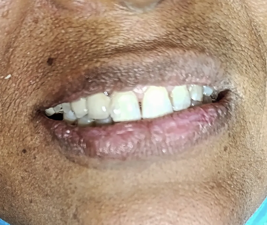 After
AfterEighteen months of avoiding photographs. A simple, well-designed bridge to end that.
The most common reason patients delay treatment for missing front teeth is not money or fear — it is the assumption that the case is somehow too complex or too expensive to address. Ms. Parveen's case is a clear example of how a single well-planned bridge can solve what feels like a much larger problem.
Ms. Parveen walked into our clinic on a cool morning in early spring. She is roughly 50 years old, a homemaker, and a quiet patient who answered our intake questions slowly and carefully — not because she was nervous but because she had thought about each one before coming in. Her chief complaint was unambiguous: “I haven't smiled in any photograph for over a year.”
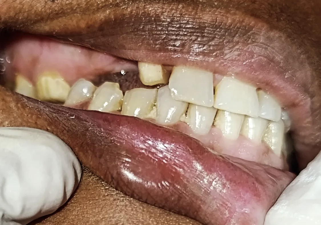
The reason was easy to see when she opened her mouth. Two of her upper front teeth were missing in the central zone of her smile, and the gap had been there for approximately eighteen months. The original teeth had been lost — she explained — to a combination of long-standing decay and a previous extraction that had not been replaced. She had been told by another clinic, around a year earlier, that she would need either implants or a complex multi-stage rehabilitation, and the quoted timeline of six months had made her postpone the decision indefinitely.
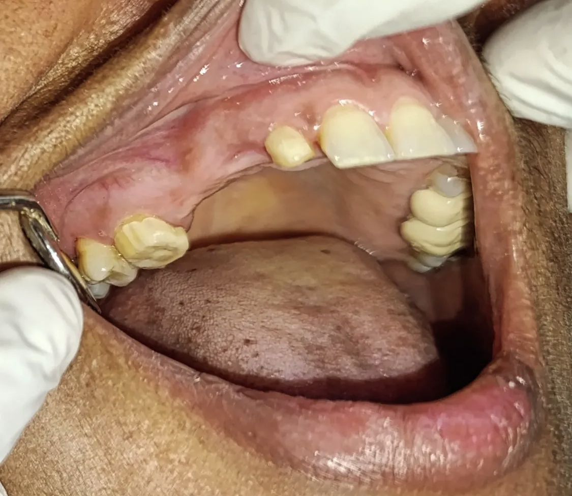
The clinical examination took 40 minutes. We charted the teeth on either side of the gap — the two lateral incisors and the two canines — and found them all to be in good condition. No decay, no large existing restorations, no periodontal pocketing greater than 3 mm. Both periapical X-rays and the panoramic radiograph confirmed that the bone support around all four potential abutments was intact. This was not a complex case. It was a routine, well-defined four-unit anterior bridge case, and it could be completed in five weeks.
We laid out two main options at the consultation. Option one was implants — two implants placed in the missing-tooth sites, with a four to six month osseointegration period before the final crowns could be loaded, and a likely need for a CBCT scan to assess bone volume, possibly a bone graft if the volume was insufficient. Option two was a four-unit anterior PFM bridge — preparation of the two laterals and two canines as abutments, a chair-side acrylic provisional bridge from the day of preparation, and a final bridge cemented within five weeks of starting.
She listened, asked sensible questions about cost and longevity for each option, and took the written treatment plan home for two days. She came back with her decision: the bridge. Her reasoning, in her own words, was that she did not want to wait six months for an implant timeline when the issue had already been there for eighteen, and the cost difference was a meaningful one in her family budget. We respected the decision and started the preparation visit the following week.
By the end of the second visit, she had a chair-side acrylic provisional bridge that looked, to a casual observer, exactly like her own teeth would have looked. By the end of the fifth visit, she had the final glazed PFM bridge cemented, the bite balanced, and the polish complete. She left with a full aftercare sheet and the next recall already in her calendar.
What made Ms. Parveen's case feel particularly satisfying was the gap between her expectation of complexity and the actual simplicity of the work. She had been told elsewhere — perhaps fairly, perhaps overcautiously — that her case would be a six-month rehabilitation involving implants, bone grafting, and a CBCT-driven surgical workflow. We did not have to do any of that. Her case was, on careful examination, a routine four-unit anterior PFM bridge case on four healthy abutments, with no need for any surgery beyond the bridge preparation itself. The decision to present it as such, rather than to upsell into the bigger plan, was the most important clinical judgment of the case.
The aesthetic result was the part that had mattered most to her, and the part we paid the most attention to. The four-unit bridge was matched against her own laterals and canines under three different lighting conditions — operatory chair light, hand-held daylight bulb, and ambient room light. We took photographs at every try-in stage and sent them to the laboratory before final approval. By the time the bridge was cemented, the shade and the contour of the new teeth were close enough to her own natural teeth that her sister — who came with her to the cementation visit and who had been instrumental in encouraging her to seek treatment — could not tell, at conversational distance, which four teeth were the new ones.
The other thing worth describing in detail is the lip-support change. Before treatment, the missing anterior teeth had been allowing her upper lip to fold slightly inward over the edentulous space. The lip support is provided by the front teeth pushing the lip outward into its natural arc. Once the new bridge was in place, the lip support was restored — and with it, the natural fullness of the upper lip and the definition of the philtrum. Subtle, but meaningful. The photographs comparing the “before” lip position to the “after” lip position show a perceptible restoration of facial proportion, and patients in this kind of case often comment that they look younger after treatment, even though no facial procedure was performed.
The maintenance plan from here is straightforward and built around routine. Floss under both pontics every night using super-floss. Brush with a soft-bristled toothbrush at the cervical margins of all four abutments. Come back every six months for a complimentary marginal check and a routine professional clean. With that schedule kept, the bridge is expected to serve her reliably for 15 years and possibly longer. At age 50, that takes her into her mid-sixties — a service life that more than justifies the conservative single-bridge approach we chose at the start.
Four findings — and an unusually clean case. Nothing complex was needed.
Not every dental problem needs a complex plan. Ms. Parveen's case is a textbook example of when a single well-designed bridge is the most efficient route to a complete solution.
Missing maxillary anterior teeth
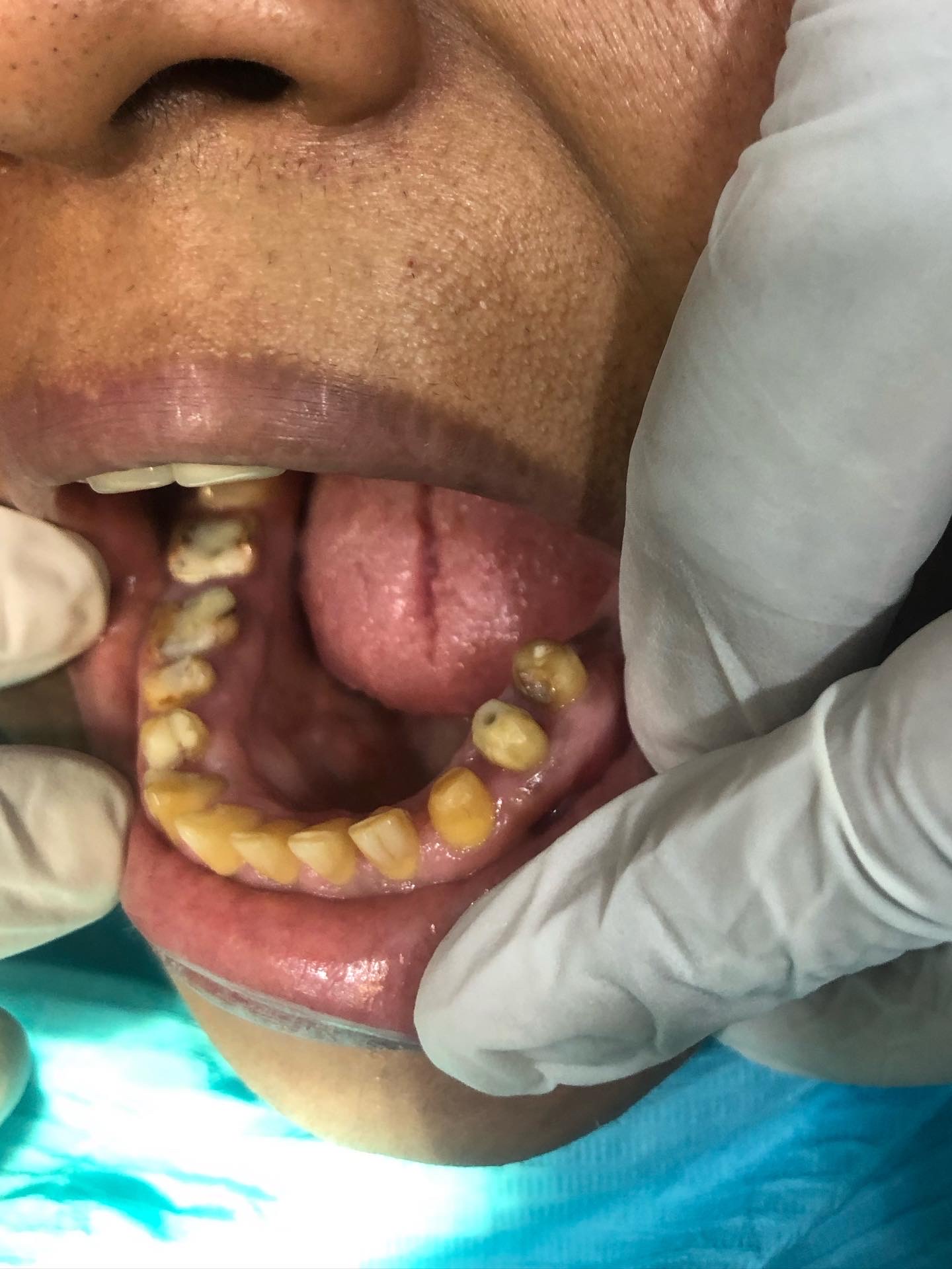
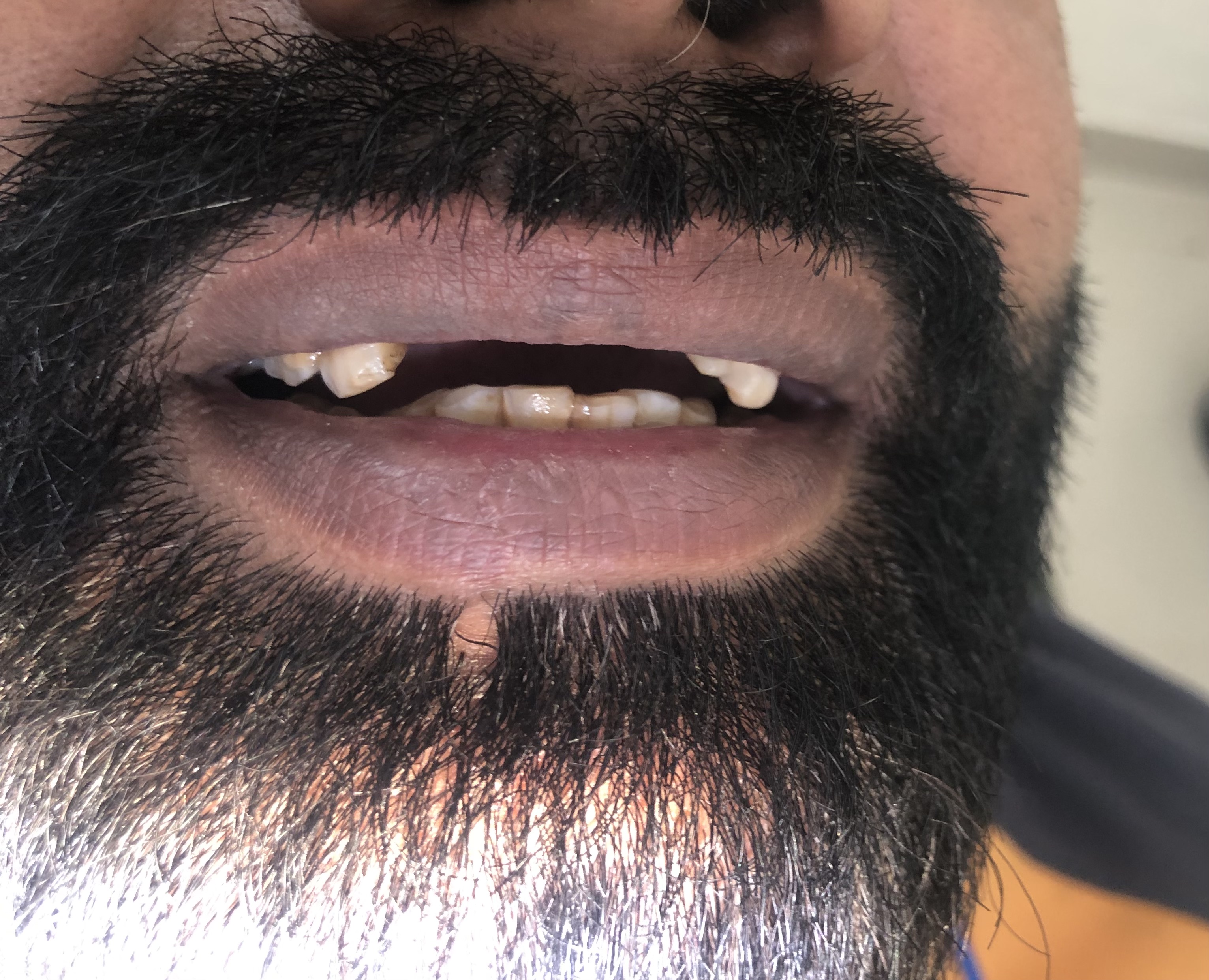
When Ms. Parveen sat in the chair and we asked her to smile, the absence was the dominant feature of her face. Two upper anterior teeth were missing in the central zone of her smile. The tooth on either side of the gap had drifted slightly inward over time, and the space had begun to close from above as well. The teeth on either side of the gap — the lateral incisors and the canines — were intact, vital, and structurally sound. They were good candidates for serving as abutments for a fixed bridge.
Compromised lip support in the upper anterior zone
When a person loses front teeth and the gap goes unrestored for any length of time, the upper lip slowly loses the support that the front teeth provided. The vermillion border curves inward, the philtrum becomes less defined, and the entire mid-face appears to age faster than the rest of the face. Ms. Parveen had been living with the gap for around eighteen months and the early signs of this lip-support loss were already visible at consultation.
Reduced functional chewing on the front teeth
Without front teeth, the patient adapts by chewing further back in the mouth and avoiding any food that requires biting or tearing with the incisors. Ms. Parveen had been doing exactly this — slicing her food into small pieces and chewing only with the back teeth. Over time this kind of compensation pattern wears the posterior teeth faster than they would otherwise wear, because they are carrying loads they were not designed for.
Healthy abutment candidates on either side of the gap
Despite the missing anteriors, the four teeth on either side of the gap were in good condition. No decay. No prior restorations. Healthy periodontal probing depths under 3 mm. Adequate bone support visible on the panoramic radiograph. They were textbook abutment candidates — strong enough to support a four-unit fixed bridge across the missing space without compromising their own long-term prognosis.
Four steps. Five visits across five weeks.
The patient had a temporary acrylic bridge that looked natural from the second visit onwards. No interval was spent in any visible “gap” state.
Consultation, photographs and bite analysis
Ms. Parveen came in for a 45-minute standalone consultation. We took six standardised intra-oral photographs, two extra-oral views with her natural smile, and a careful study of how her lip moved over the missing space when she spoke and when she laughed. We then took two periapical X-rays of each potential abutment tooth plus a single panoramic radiograph. The plan and a written quote were placed in her hand before she left.
Visit 1 · ~ 45 minTooth preparation and provisional bridge
At the second visit we numbed both quadrants where the abutment teeth were located, and prepared the four abutment teeth to receive porcelain-fused-to-metal crowns. The preparations were conservative — a 1.2 mm reduction labially, 1.5 mm incisally, and a chamfer finish line at the gum level. A digital scan and a chair-side acrylic provisional bridge were made so she walked out of the appointment with a fully shaped temporary that looked like her own teeth.
Visit 2 · ~ 90 minBridge fabrication and try-in
The PFM bridge was returned from the laboratory two weeks later. We tried in the metal substructure first, then the biscuit-bake porcelain, then the final glazed bridge — three stages, each of which gave her the chance to look in the mirror and approve the next step. At each stage we checked the marginal fit, the occlusion in centric and protrusive movements, the proximal contacts with the adjacent teeth, and the shade match against her own laterals and canines.
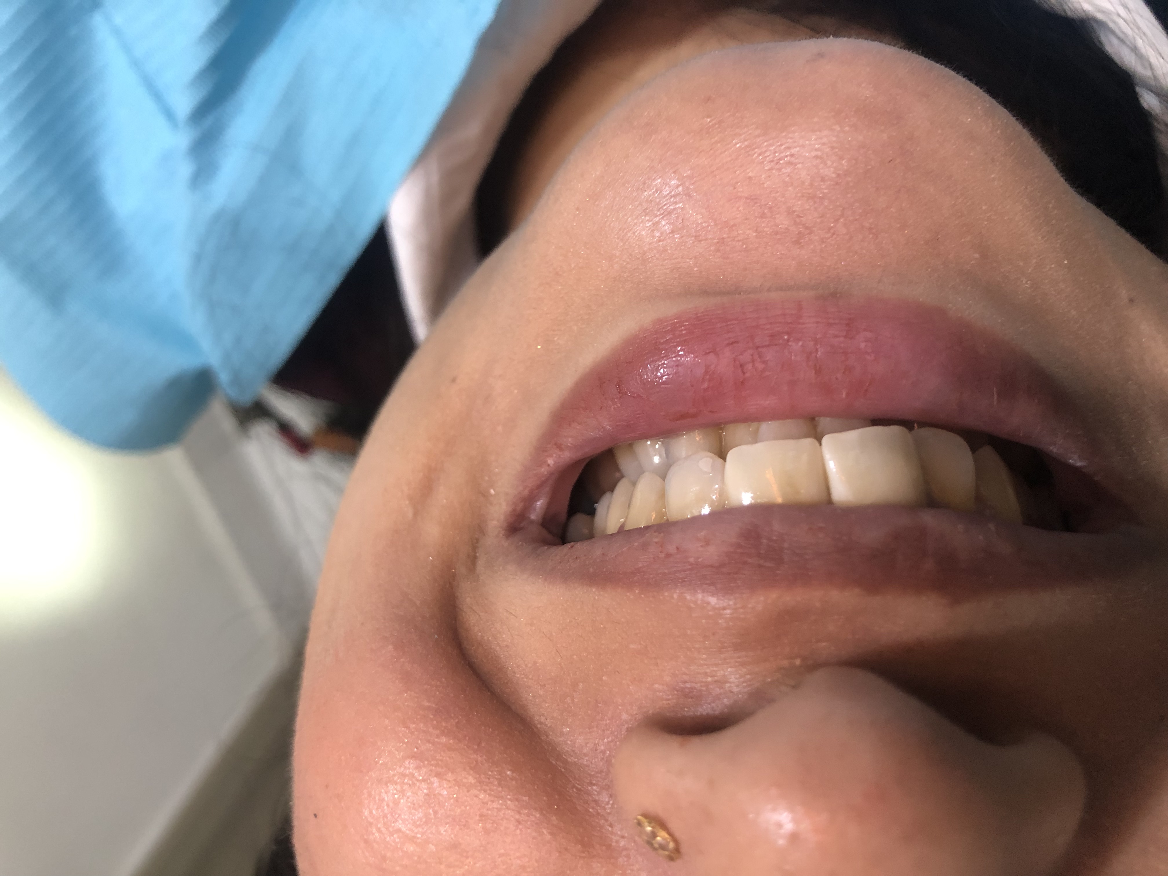
Visits 3-4 · across 3 weeksFinal cementation and polish
The bridge was cemented with a self-adhesive resin cement. We removed the excess cement under magnification, polished the labial surface to a high gloss, and checked the bite one last time. She left with a written aftercare sheet, a sample bottle of chlorhexidine mouthwash for the first ten days, and her next visit pencilled in for the one-week review.
Visit 5 · ~ 50 minSame patient. Eighteen months of absence, reversed in five weeks.
Drag the divider to compare. The "before" image is from the first consultation; the "after" image is from the day of final cementation.
BeforeAfterWhy a bridge can be the right answer even when implants are available.
Three things determine whether a missing-tooth case is best treated with a bridge or with implants. All three favoured a bridge in Ms. Parveen's case.
Adjacent abutment teeth condition
When the teeth on either side of a missing-tooth gap are already heavily restored — large fillings, prior crowns, or fractures — preparing them for a bridge is essentially using the same teeth that would have needed treatment anyway. When the adjacent teeth are intact and healthy, the decision is more nuanced. For Ms. Parveen, the four abutment candidates were all unrestored, vital, and structurally sound — strong starting points for either approach.
Bone volume in the missing-tooth region
Implants require adequate bone at the site of placement. After eighteen months of missing teeth, Ms. Parveen had already lost a small amount of vertical and horizontal bone in the edentulous region — predictable resorption that begins within weeks of extraction. An implant plan would have required a CBCT scan and possibly a bone-graft procedure before any implants could be placed. A bridge avoided that surgical step entirely, because the bone in the missing-tooth region does not need to support the bridge — the abutment teeth do.
Time, cost and predictability for the specific patient
For a 50-year-old patient who had already waited eighteen months and wanted a fast, predictable solution within a clear budget, a four-unit anterior bridge offered all three. The implant option, while excellent in its own right, would have meant another six-month wait and roughly twice the cost. The choice was hers to make after we walked through both honestly. The bridge route was the right one for her circumstances.
Five questions we hear at every anterior bridge consultation.
These are the worries we heard from Ms. Parveen, and the worries we hear from most patients considering a fixed bridge for missing front teeth.
Will the bridge teeth look the same colour as my natural teeth?+
For most patients, yes — the bridge teeth can be shade-matched extremely closely against the adjacent natural teeth. The key is the careful shade study we do at the chair before sending the case to the laboratory. We compare against three different shade tabs under both daylight and clinic light, photograph each comparison, and send the photographs to the technician along with the impression.
Modern porcelain-fused-to-metal crowns are layered with three or four shade gradients from cervical to incisal, plus a translucency profile tuned to imitate enamel. At try-in, we check the match against the patient's actual lip and skin tone — not just against the shade tabs. If anything looks off, the case goes back to the laboratory before final cementation.
For Ms. Parveen, the four-unit bridge was matched against her natural laterals and canines, and the match was checked at three try-in stages before cementation. At six months, she has not had a single comment from anyone in her family or social circle about the bridge teeth looking different. That is the bar we aim for.
How long does an anterior PFM bridge last?+
A well-made PFM bridge on healthy abutment teeth with disciplined home care lasts 12 to 20 years in the published literature. Our own clinic experience has many bridges past the 15-year mark with no marginal breakdown and stable surrounding gums.
The single biggest predictor of bridge longevity is plaque control at the cervical margin and under the pontics. That is why the aftercare regimen — flossing under the pontics every night, soft brushing at the margin, six-monthly recall — matters more than the brand of porcelain or the cement used.
For Ms. Parveen at age 50, an expected 15-year service life would take her bridge into her mid-sixties. At that point, depending on the state of the abutment teeth, the bridge can either be re-made (if the abutments are still healthy) or transitioned to an implant-supported bridge (if any of the abutments has failed). The plan is built with both options open.
Why not implants instead of a bridge?+
Implants are an excellent option for missing teeth and we present them at every consultation involving a gap that could be restored either way. For Ms. Parveen we walked through the trade-offs honestly.
Implants would have meant a longer total timeline (typically 4 to 6 months for osseointegration before final loading), a higher cost (roughly two to three times the cost of the bridge), and at least one surgical procedure with a 4-month healing window. They also require adequate bone volume in the missing-tooth region, which we would have needed to confirm with a CBCT scan and possibly a bone graft if the bone was insufficient.
The bridge option offered a faster timeline (about 5 weeks total), a substantially lower cost, no additional surgery, and a predictable 15-year service life. The abutment teeth on either side of the gap were both healthy and would not be required to do anything they were not designed for. For her case, the bridge was the right balance of cost, time, and longevity. She made the decision after taking the plan home for two days.
Will the abutment teeth on either side be weakened by the bridge?+
The honest answer is that an abutment tooth is a tooth that has been prepared — meaning some enamel and a small amount of dentine have been removed to make room for the bridge crown. That preparation does reduce the structural reserve of the tooth, particularly compared to its untouched state.
In practice, however, a properly prepared and crowned abutment tooth is significantly stronger than the same tooth with a large filling or visible crack would have been. The crown encloses the cusps and the proximal walls in a single rigid shell, which actually distributes biting forces more evenly than a natural tooth with a large internal restoration.
For Ms. Parveen the four abutments were all unrestored, intact, vital teeth — about as strong a starting point as we ever see. The bridge preparation reduced their structural reserve modestly, but the new crown protection more than compensates. We expect those abutments to outlast the bridge itself.
What happens at the six-month and twelve-month recall visits?+
Every patient with a fixed bridge at our clinic returns for a complimentary marginal check at six months, and a fuller routine recall at twelve months. The structure of these visits is standardised so that any developing problem is caught at the smallest possible intervention point.
At the six-month visit, we re-photograph the bridge under the same lighting as the cementation appointment, probe the gum at six points around each abutment, run floss under each pontic to confirm the contour is still right, and inspect the cervical margins for any developing staining or marginal breakdown. The visit takes about 20 minutes. If everything is healthy, there is no further work to do at that appointment. If we detect any early sign of trouble, we discuss it then and there and plan the small intervention needed.
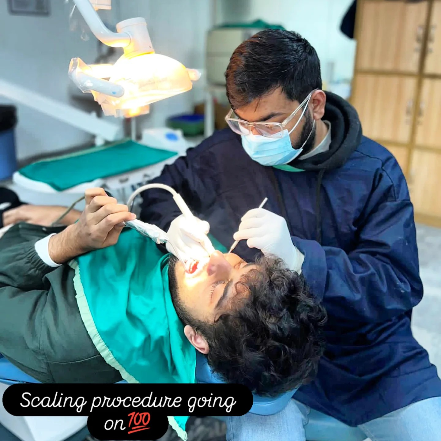
At the twelve-month visit, we repeat all of the above plus a routine professional clean of the entire mouth. Plaque has a way of accumulating in places the patient cannot quite reach — particularly at the embrasures of the bridge — and a careful scaling once a year keeps the long-term margins clean. If a bite-wing X-ray of any abutment looks indicated, we take one. The total visit time is typically 45 to 60 minutes.
For Ms. Parveen, the six-month and twelve-month reviews both confirmed that her home care was holding the result. No early marginal staining. No bleeding on probing. No food impaction at any of the pontic embrasures. We have her booked for the second-year check in approximately six months from the time of this case file.
How much does a four-unit anterior bridge cost?+
Our PFM bridge fees are quoted per unit and include the abutment preparation, the laboratory work, all try-in visits, and the final cementation. For a typical four-unit anterior bridge at our clinic, the total cost ranges from PKR 60,000 to PKR 110,000 depending on the laboratory used and the number of try-in stages required.
The quote is always written and itemised before any work begins. There are no additional charges for the consultation, the diagnostic photographs, or the six-monthly recall checks during the first year. A routine scaling at the same visit, if needed, is charged separately at our standard scaling fee.
For Ms. Parveen, the four-unit anterior bridge was completed within a single, all-inclusive fee. No add-ons appeared on the final invoice that were not on the original written quote.
The follow-up visits.
An anterior bridge is finished six months after cementation — once the gum has fully matured around the new margins and the patient has lived with the new bite long enough to flag anything that does not feel right.
Routine bite check and marginal probing. We polished a single high spot we had detected on the right-canine abutment. She reported zero sensitivity and was eating on the front teeth comfortably for the first time in over a year.
The cervical margins of all four abutments had matured. No bleeding on probing at any site. The pontic-pink-gingiva relationship looked natural and was photographing well.
Complimentary six-month marginal check. All margins sealed. No marginal staining. Probing depths under 3 mm everywhere. She demonstrated her flossing technique under the pontics — perfect.
Dr. Mian Momin Ahmad
“Cases like Ms. Parveen's are reminders that the right plan is almost always the smallest one that actually solves the problem. A four-unit anterior bridge on four healthy abutment teeth is not a glamorous procedure, but it is a deeply useful one. The patient came in having postponed treatment for eighteen months because a previous clinic had quoted her something larger. Five visits later, she had her smile back.”
Six habits that hold the result for 15 years.
Anterior bridges last as long as the gum and the abutment teeth stay healthy. These six habits are what we asked Ms. Parveen to commit to.
Floss under the pontics every night
A four-unit bridge has two pontics — the tooth-shaped sections that fill the gap. The undersurface of each pontic sits just over the gum and is the single most common site for plaque retention in any bridge. Super-floss or a floss-threader slid under each pontic once a day, takes ten seconds per pontic. Done every night, this is the habit that determines whether the bridge lasts twelve years or twenty.
Use a soft toothbrush at the cervical margin
The cervical margin where the bridge meets natural tooth is the second most common site for problems — most often gum recession driven by aggressive brushing. A soft-bristled brush, small circular motions, two minutes morning and night, is exactly the right cadence. We sized Ms. Parveen up for an Oral-B Sensitive head.
Don't bite directly into apples or hard naan crust
A modern PFM bridge is strong enough for normal eating, but biting directly into hard or dense foods with the bridge units stresses the cement margins in a way the bridge was not designed to absorb. We asked her to slice apples into small pieces and chew with her back teeth, and to keep hard naan and chicken bones away from the front bridge altogether.
Come in if the bite ever feels off
Over time, the occlusion on a bridge can shift slightly — usually because of wear on opposing teeth, not because of the bridge itself. The fix is a five-minute occlusal adjustment with articulating paper at a routine visit, not a re-do of the bridge. We asked her to flag any new sense of "the teeth meeting wrong" the moment she noticed it.
Avoid whitening toothpastes for the first three months
Whitening toothpastes are usually mildly abrasive and can scratch the surface glaze of a freshly polished PFM crown. For the first three months while the cement margin is still maturing, a regular fluoride toothpaste — Sensodyne Sensitive or Colgate Total — is the right choice. After three months she can resume whatever paste she prefers.
Six-monthly recall, every time
Every bridge patient at our clinic comes back at six months for a complimentary marginal check. We photograph the cervical margin under magnification, probe the gum at six points around each abutment, and remove any plaque that has gathered where her floss could not quite reach. Twenty minutes per recall, and it catches almost every long-term problem at the small-intervention stage.
Every month a front tooth is missing, the case changes shape.
Patients who delay restoring missing anterior teeth often assume the case will be the same case in a year. It rarely is. Bone resorbs at the extraction site, the adjacent teeth drift, the lip-support loss becomes harder to reverse, and the gum architecture changes in ways that the eventual prosthesis has to compensate for.
Ms. Parveen came in at the eighteen-month mark. Her case was still straightforward. The earlier the intervention, the simpler the eventual restoration.
More on bridges and what they replace.
Three more crown and bridge patients.
Every case in this archive is a real Odonto patient with their written consent.
Missing front teeth? Let's plan it properly.
The first 15 minutes are free. We will examine the gap, examine the teeth on either side, and put a written plan with both bridge and implant options in your hand. No pressure to commit the same day.