How a small cavity, caught at the right time, became a single-visit filling — Qari Sultan's story.
Qari Sultan Mahmood came to our Engineers Town clinic with a pattern of pain almost every dentist recognises immediately — short, sharp, food-triggered, gone within ten minutes. This is the case file for the single-visit laser-assisted Class II composite filling that resolved it, and the reason why coming in at this stage of a cavity is one of the most efficient moments in dentistry.
 Before
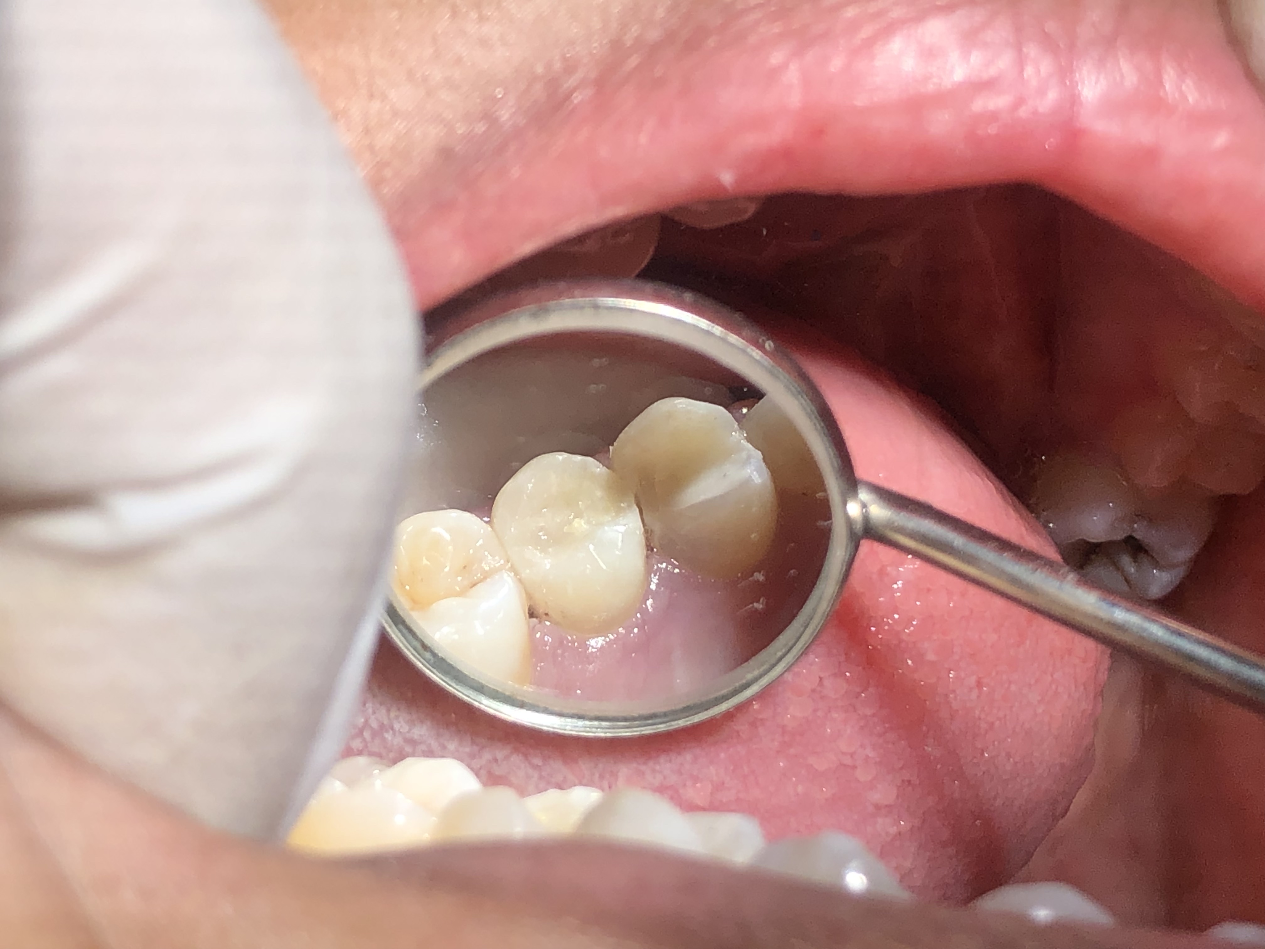
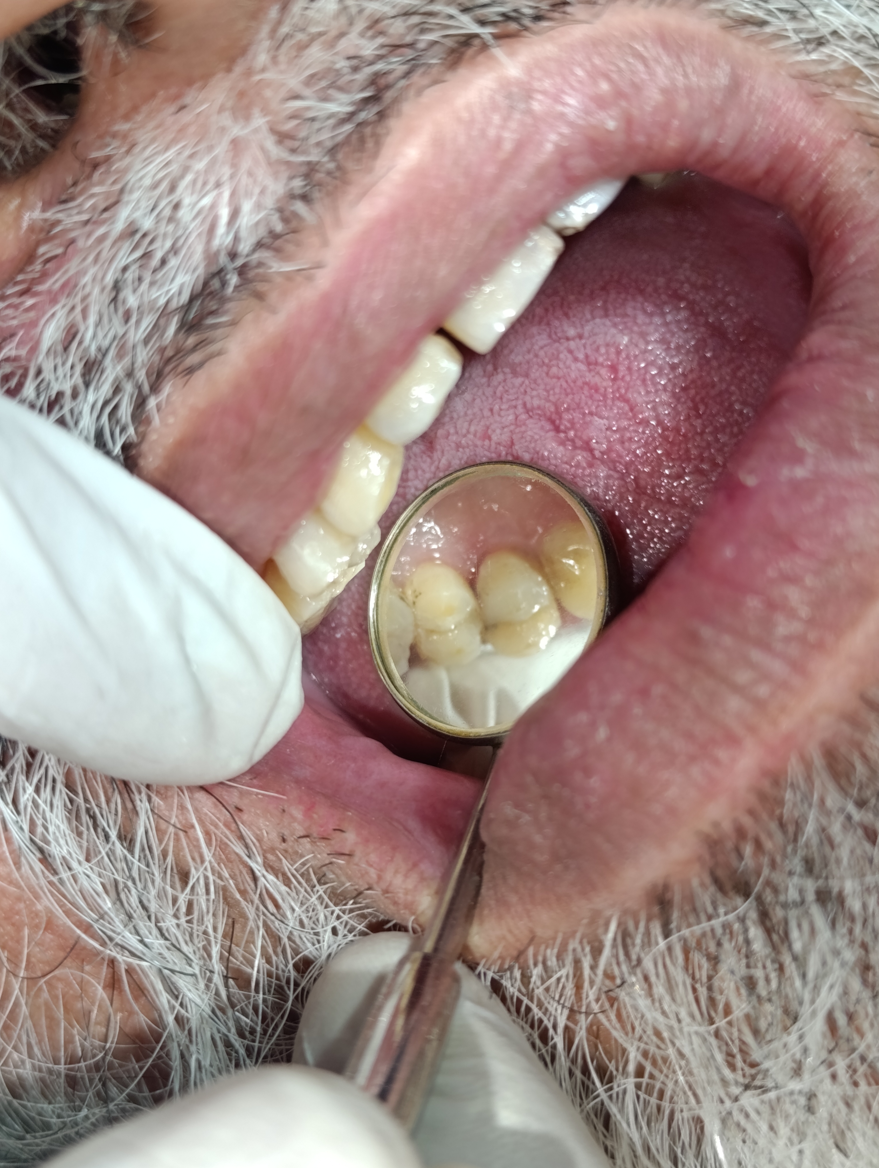 After
AfterA predictable pattern. A predictable fix.
Pain that comes on while eating and ends within ten minutes is one of the most informative things a patient can tell a dentist. It usually has a single explanation, and that explanation is usually fixable in one visit.
Qari Sultan Mahmood came in to our clinic on a Tuesday afternoon. He is a working adult patient who had been managing — but not enjoying — a recurring pattern of pain during meals for around two months. He described it precisely: it came on as he chewed, lasted for the length of a few bites, peaked sharply once or twice in the middle of a meal, and then subsided within about ten minutes of finishing the food. He had no spontaneous pain. He had no night pain. He had no pain at rest at all.
That description, almost more than any other piece of information a patient can give, is diagnostic. Stimulus-provoked pain that ends shortly after the stimulus ends is the textbook description of reversible pulpitis — a condition in which the pulp of a tooth is irritated by a nearby cavity but has not yet been damaged irreversibly. The treatment is to remove the cause (the cavity) and the symptoms resolve completely. The treatment is a filling, not a root canal.
The clinical examination took 15 minutes. We took a focused intra-oral photograph of the affected quadrant, a periapical X-ray, and performed a cold-test on the affected tooth as well as the two adjacent teeth (to rule out referred pain coming from a neighbour). The X-ray showed the lesion clearly — a small triangular dark zone at the proximal contact point of one of the posterior teeth, extending inward toward the enamel-dentine junction but not yet reaching the pulp. The cold-test response was sharp, brief, and ended cleanly. Everything fit.
We walked Qari Sultan through what we were going to do, with the X-ray on the screen between us. The cavity would be cleaned out under local anaesthesia, the floor disinfected with a soft-tissue laser as we worked, and the tooth restored with a tooth-coloured composite resin. The bonding would be done under a rubber dam to keep the field dry — a small detail that dramatically improves how long a filling lasts. The whole procedure would take about 70 minutes of chair time, and he would be able to eat normally the same evening.
He signed the consent form, we numbed the quadrant, and we began.
The decision to use a laser-assisted technique for a case like Qari Sultan's deserves a note of explanation. Many patients assume the laser does the actual cavity preparation — replacing the dental drill — and that this is somehow softer or less invasive. That is not how it works. The rotary bur is still the instrument that removes the bulk of the carious tissue. The laser is a complementary tool that we pass over the cavity floor between the rotary stages, disinfecting the dentine surface and reducing the bacterial load before the bonding adhesive is applied. The published evidence for laser-assisted disinfection in cavity floors is modest but consistent — a small reduction in post-operative sensitivity, and a small improvement in the long-term seal of the restoration. Neither is dramatic, but together they justify the use of the laser as an adjunct in cases like this.
The most under-appreciated aspect of a Class II composite filling is the matrix system used to recreate the proximal contact. For Qari Sultan's tooth, we used a sectional matrix — a small curved metal band held in place by a flexible ring that pushes the band outward against the proximal wall, recreating the natural bulge of a healthy contact point. The alternative is a circumferential matrix that wraps around the entire tooth. Sectional matrices produce snap-fit proximal contacts with the same tightness as natural teeth; circumferential matrices are easier to place but tend to produce slightly looser contacts. The difference shows up at the six-month recall: patients with sectional-matrix restorations rarely report food impaction at the contact, while patients with circumferential-matrix restorations sometimes do. Small details, large long-term consequences.
One final detail worth recording. The rubber-dam isolation we used for this filling is the single most important determinant of how long the restoration will last. The composite resin we used bonds to the dentine through an adhesive layer that is acutely sensitive to moisture contamination. A drop of saliva on the bonding surface during placement compromises the long-term strength of the adhesion. Rubber-dam isolation eliminates that risk completely. Bonded restorations placed under rubber dam outlast restorations placed without it by several years in every published comparison. For a 70-minute case, the extra five minutes spent placing the rubber dam at the start is the most cost-effective decision in the entire procedure.
By the end of the appointment, Qari Sultan was sitting up with the dam removed, the bite checked, and the proximal contact confirmed by floss. He drank a glass of water, washed his hands, and walked out the same afternoon. He messaged that evening to say he had eaten dinner normally, with no pain on either side. The 70-minute procedure had ended a pattern of pain that had been quietly disrupting his meals for two months.
Four findings — all consistent with one diagnosis.
The skill in a case like this is not in performing the filling. It is in confirming, before any treatment begins, that a filling is in fact the right answer — and not a sign of something deeper.
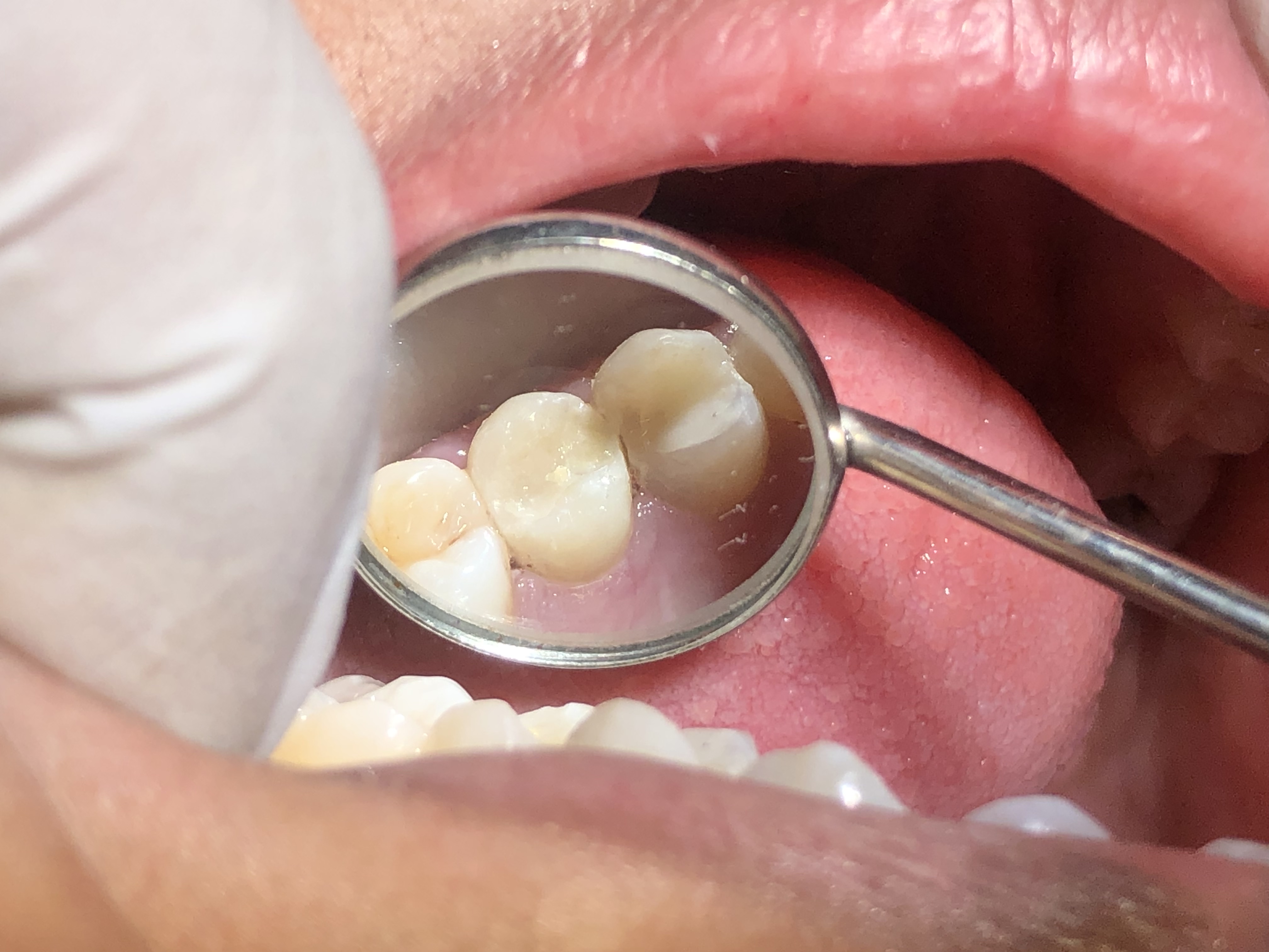
Proximal Class II caries on a posterior tooth
When we shone the operating light into Qari Sultan's mouth and examined the posterior teeth one by one with a fine dental explorer, we found the source of his eating pain — a small but definite carious lesion on the proximal surface (the side that faces the neighbouring tooth) of one of his molars. The lesion was visible on the radiograph as a triangular dark zone extending from the contact point inward toward the enamel-dentine junction. This is the classic appearance of an early Class II carious lesion: the kind that has been quietly developing for months and only becomes symptomatic when the patient starts catching food in it.
Symptomatic reversible pulpitis — pain that ends shortly after eating
The pattern Qari Sultan described was extremely useful diagnostically. The pain came on while he was chewing food, peaked sharply for a few seconds, then subsided within about ten minutes of stopping. There was no spontaneous pain, no night pain waking him up, no pain that lingered for hours after the stimulus had been removed. That pattern — pain provoked by a stimulus, ending shortly after the stimulus stops — is the textbook description of reversible pulpitis. The pulp is irritated but not damaged. Treat the cause (the cavity) and the symptoms resolve completely.
No deeper pulpal involvement on cold testing
We performed a cold-stimulus test on the affected tooth using a small refrigerant spray on a cotton pellet. The response was sharp, brief, and ended cleanly within four to five seconds of removing the stimulus. This is exactly the response we hope to see — it confirms the pulp is alive, the nerve is functional, but the inflammation is mild and reversible. A response that lingers for more than ten or fifteen seconds would have suggested irreversible pulpitis and a likely root canal. The crisp short response we got told us a filling would resolve the case.
No periapical changes — healthy bone around the root
The periapical radiograph showed completely normal periapical bone around the root tips of the affected tooth. There was no widening of the periodontal ligament space, no peri-apical radiolucency, and no signs of infection extending into the surrounding bone. The bony surroundings were quiet. Combined with the clinical findings, this confirmed that the entire problem was confined to the cavity itself. A single restorative visit was all that was needed.
Four phases. All in one sitting. About 70 minutes total.
Every phase was explained before it happened. He was able to look at the procedure on the screen above the chair at any point.
Examination, photographs and X-ray confirmation
We took a focused intra-oral photograph of the affected quadrant, a periapical X-ray to confirm the depth of the lesion, and a cold-test response check on the tooth and on the two adjacent teeth (to rule out referred pain). We explained the diagnosis to Qari Sultan with the X-ray on the screen — the cavity was visible as a small dark wedge between his molar and the neighbouring tooth — and we walked through the plan.
Phase 1 · ~ 15 minLocal anaesthesia and rubber dam isolation
A buccal infiltration of 4% articaine numbed the quadrant within five minutes. We placed a rubber dam over the affected tooth and the two adjacent teeth, which gave us a completely dry isolated working field. Rubber dam isolation is one of those details that does not feel exciting to a patient but is the single most important determinant of how long a filling lasts — bonding in a saliva-contaminated field fails far sooner than bonding in a dry, isolated field.
Phase 2 · ~ 10 minLaser-assisted caries excavation
We used a soft-tissue diode laser to assist with the caries excavation phase. The laser disinfects the floor of the cavity as the rotary bur removes the soft, infected dentine — reducing the bacterial load at the pulpal floor and reducing the risk of post-operative sensitivity. We checked the cavity floor with a caries-detector dye to ensure all infected dentine had been removed before proceeding to bonding. The remaining sound dentine was etched with phosphoric acid, rinsed, and prepared for the adhesive.
Phase 3 · ~ 20 minComposite layering, contour and polish
We placed a sectional matrix system around the affected tooth to recreate the proximal contour. The composite was placed in 2 mm horizontal layers, each light-cured for 20 seconds before the next was added. Once the cavity was fully built up, we removed the matrix, checked the proximal contact with floss (snap-fit, exactly the right tightness), and contoured the occlusal anatomy with a fine diamond bur. A final polish with a sequence of rubber polishing wheels brought the surface to a high gloss.
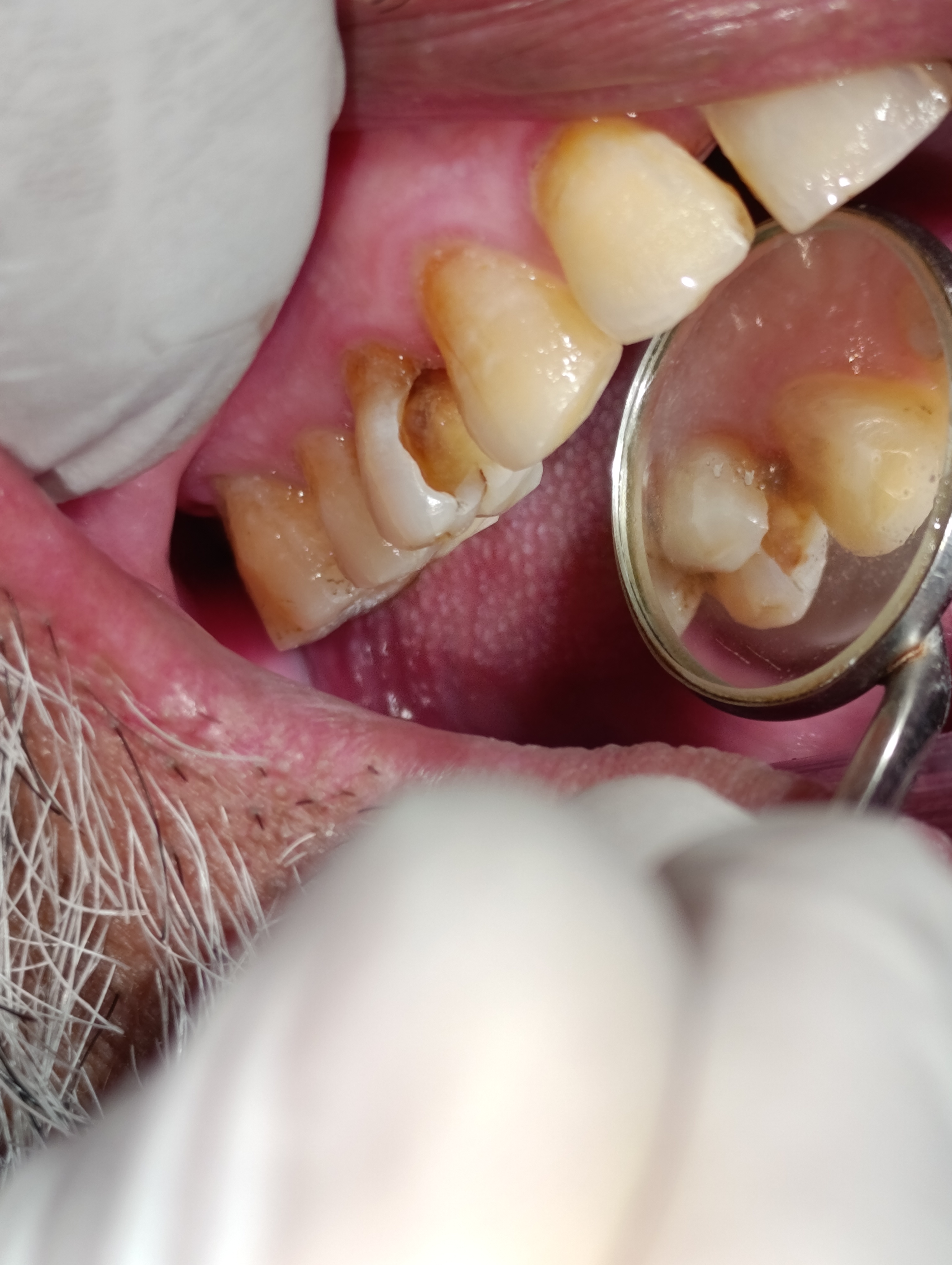
Phase 4 · ~ 25 min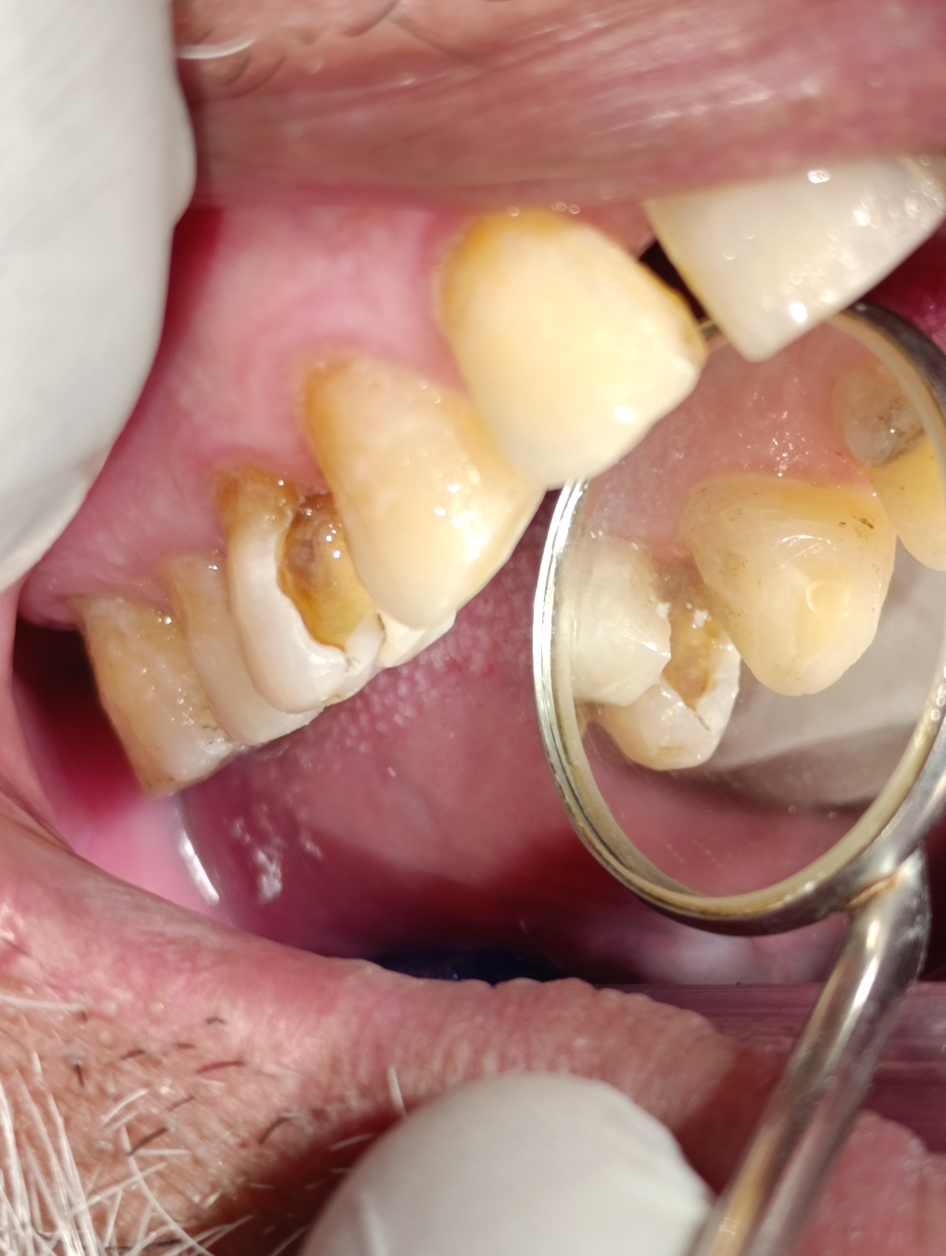 During — cavity preparation
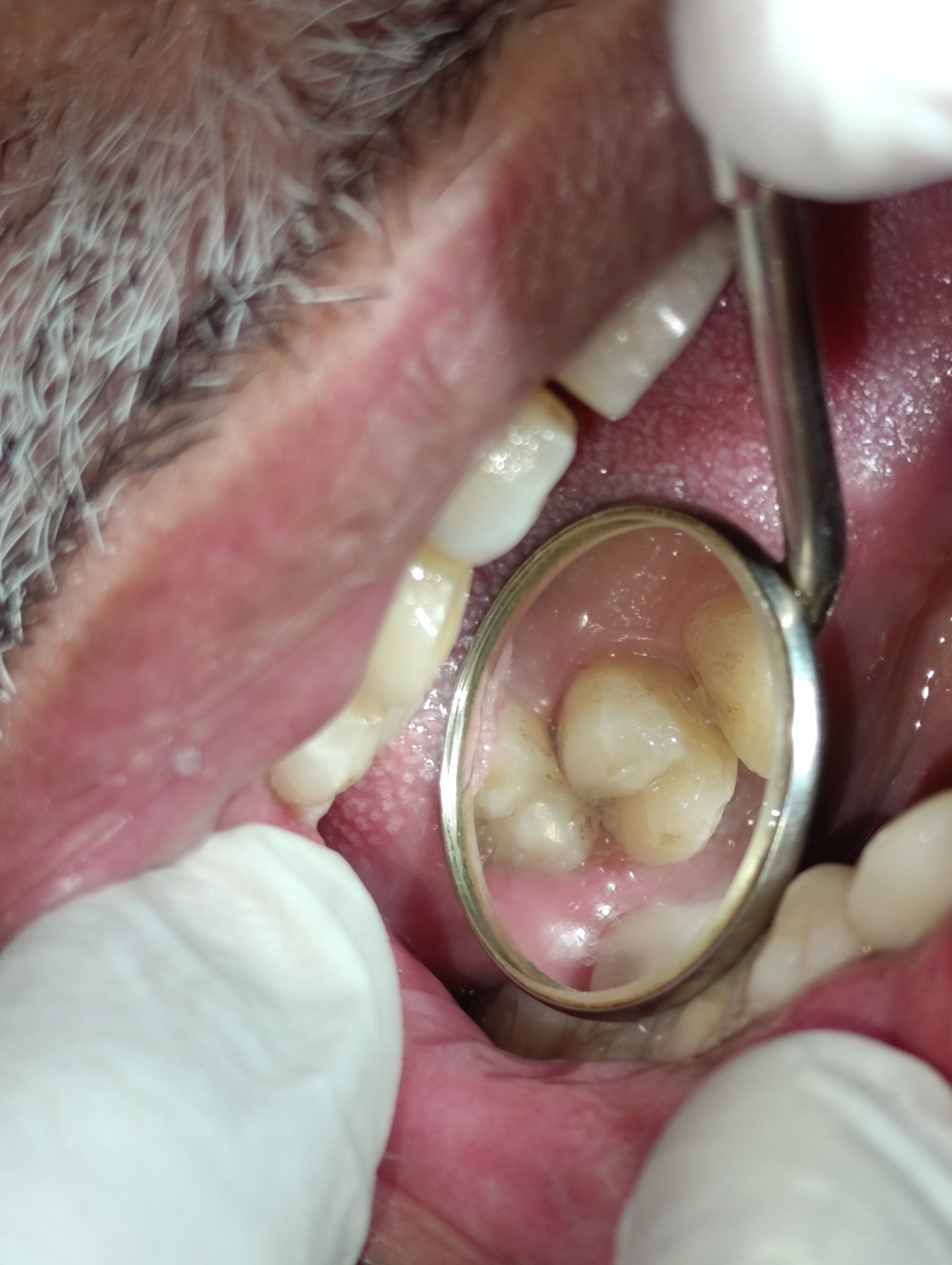 During — composite layering
Same patient. Same tooth. Same day.
Drag the divider across the photo to compare. The "before" image is from the diagnostic visit; the "after" image is the completed restoration on the same day.
BeforeAfterWhy a Class II composite is harder than it looks.
A simple posterior filling sounds routine. The technical details that determine whether the same filling lasts five years or fifteen are anything but. Three of them are worth knowing about.
The proximal contact has to be exactly the right tightness
If the contact between the new filling and the neighbouring tooth is too loose, food packs into the embrasure at every meal and the cavity recurs within years. If the contact is too tight, the patient cannot floss properly and plaque accumulates at the margin. The sectional-matrix system we use creates a contact that is snap-fit — exactly the same tightness as the contact between two natural teeth. This is the single most under-rated detail of a Class II filling.
A dry working field changes the failure rate dramatically
Composite resin bonds to dentine through an adhesive layer that is acutely sensitive to moisture contamination. A drop of saliva on the bonding surface during placement compromises the long-term strength of the adhesion. Rubber-dam isolation — a small sheet of latex stretched over the working tooth — eliminates this risk completely. Bonded restorations placed under rubber dam outlast restorations placed without it by years, in every published comparison.
Composite is layered in 2 mm increments, not packed in one piece
Composite resin shrinks slightly as it polymerises. If a large cavity is filled in a single bulk increment, the shrinkage forces concentrate at the bonded interface and can pull the filling away from the tooth wall, creating a microscopic gap. Filling in 2 mm horizontal layers, with each layer cured for 20 seconds before the next is added, distributes the shrinkage across many smaller, lower-stress increments. The cavity at the end is the same size; the bonded interface is significantly stronger.
Five questions we hear at every filling consult.
These are the worries we heard from Qari Sultan and the worries we hear from most adult patients considering a posterior filling. Tap any one to read the long answer.
Will the filling change colour or stain over time?+
Modern composite resins are extremely colour-stable and rarely stain visibly within the first 8 to 10 years of service. The earlier generations of composite — used a decade or two ago — were more prone to picking up tea and coffee stain over time, which is the source of the common worry. The materials we use today are different.
That said, the proximal surface of the filling is also next to the embrasure where food contacts collect daily, and aggressive consumption of staining foods — paan, gutka, very strong tea or coffee — can eventually mark the labial border of any restoration. For a patient who consumes any of those routinely, we ask them to flag it at the consultation so we can choose a slightly higher-shade-stability composite at the outset.
For Qari Sultan, who is not a paan or gutka user, we expect the shade to remain stable through the full service life of the restoration.
How long does a Class II composite filling last?+
Class II composite restorations on adult posterior teeth have published service lives ranging from 8 to 15 years, with the median typically around 10 to 12 years. The single biggest determinant of how long any particular filling lasts is the quality of the bonding at the proximal margin, which depends on three things: the dryness of the field at the time of bonding (we use rubber dam for every Class II), the technique used to recreate the proximal contour (we use sectional matrix systems), and the home-care habits of the patient.
For a patient like Qari Sultan with a healthy mouth, good baseline hygiene, and disciplined six-monthly recalls, a 12-year service life is realistic. After that, the most common outcome is that the filling is replaced — not that the tooth is lost.
Why did you use a laser? Is that necessary or just marketing?+
The soft-tissue diode laser we use during the caries excavation phase has two specific functions that justify its use. First, it disinfects the floor of the cavity as the rotary bur exposes the dentine — reducing the bacterial load at the pulpal floor and reducing the risk of post-operative sensitivity. Second, in the rare situation where a small amount of soft tissue (gum) has overgrown into a deep proximal cavity, the laser can be used to contour that tissue without bleeding, which keeps the working field dry for bonding.
It is not marketing. The published evidence for laser-assisted disinfection in cavity floors is solid, and the small reduction in post-operative sensitivity is one of the reasons our patients tolerate Class II fillings particularly well. But it is also not magic — the laser does not replace the rotary instruments, it complements them.
For Qari Sultan, the use of the laser was part of the reason he was able to walk out of the clinic with a sealed restoration and zero post-operative sensitivity beyond the first 24 hours.
Could this have turned into a root canal if I had waited longer?+
Yes — and this is probably the most important thing to understand about reversible pulpitis. The condition is reversible only as long as the underlying cause (the cavity) is addressed before the pulp transitions from reversibly inflamed to irreversibly inflamed.
In Qari Sultan's case, the cold-test response was sharp and brief, which is the signature of mild, reversible inflammation. If he had waited another six to twelve months with the cavity progressing, the inflammation would likely have advanced. At that point the cold-test response would have become lingering rather than brief — the signature of irreversible pulpitis — and the treatment would have moved from a filling to a root canal.
The window between “a filling can fix this” and “you need a root canal” is sometimes only a few months. Coming in for the filling at the reversible stage is one of the few moments in dentistry where the patient's timing really matters.
What is the difference between a Class II filling and an MOD filling?+
A Class II filling is a tooth-coloured restoration that involves one proximal surface of a posterior tooth plus the adjacent occlusal surface. The cavity preparation crosses from the chewing surface of the tooth into the side facing the neighbouring tooth, and the filling rebuilds both surfaces. Qari Sultan's case was a single-surface Class II — only one proximal side was involved.
An MOD filling, on the other hand, involves both proximal surfaces of the same tooth — the mesial side (toward the front of the mouth), the occlusal surface, and the distal side (toward the back). MOD restorations are technically more demanding because two separate proximal contacts must be re-created, and they remove more tooth structure than a single-surface Class II. They are usually placed when bilateral caries develops on the same tooth.
Both types of restoration use the same composite resin material, the same bonding technique, and the same sectional-matrix approach to recreating the proximal contour. The difference is in scope, not in technique. The choice between them is determined by the extent of the caries, not by patient preference.
For a routine Class II case like Qari Sultan's, the procedure is well within a 70-minute single-visit appointment. For an MOD case, the appointment typically runs 90 minutes because of the extra time needed to recreate the second proximal contact and to manage the larger cavity volume.
How much does a Class II composite filling cost?+
Our composite filling fees depend on the size of the cavity and the number of surfaces involved. A Class II restoration like Qari Sultan's — one proximal surface plus the occlusal — is a routine intermediate-sized filling. The fee includes the consultation visit if it leads directly to treatment, the radiograph, the local anaesthesia, the laser-assisted excavation, the rubber-dam isolation, the composite restoration, and the polish.
The total is quoted in writing at consultation and is included in the procedure fee. There are no separate charges for the cold-test, the X-ray, or the laser use. If a second filling is needed in the same visit on a neighbouring tooth, it is charged at our second-filling rate, also quoted in advance.
For Qari Sultan, a single Class II filling completed the case. No additional charges were added at any point.
The follow-up visits.
A single-visit filling is followed by a single short review visit at the one-week mark, and then by routine six-monthly recall. Here is how Qari Sultan's follow-up went.
He messaged on WhatsApp the evening of the treatment to confirm he had eaten dinner normally on the opposite side. No new pain. The numbness had worn off cleanly within four hours.
A short 10-minute visit to check the bite with articulating paper. One small high spot detected and adjusted in 30 seconds. The proximal contact was snap-fit. No marginal staining. He had resumed full normal eating on both sides.
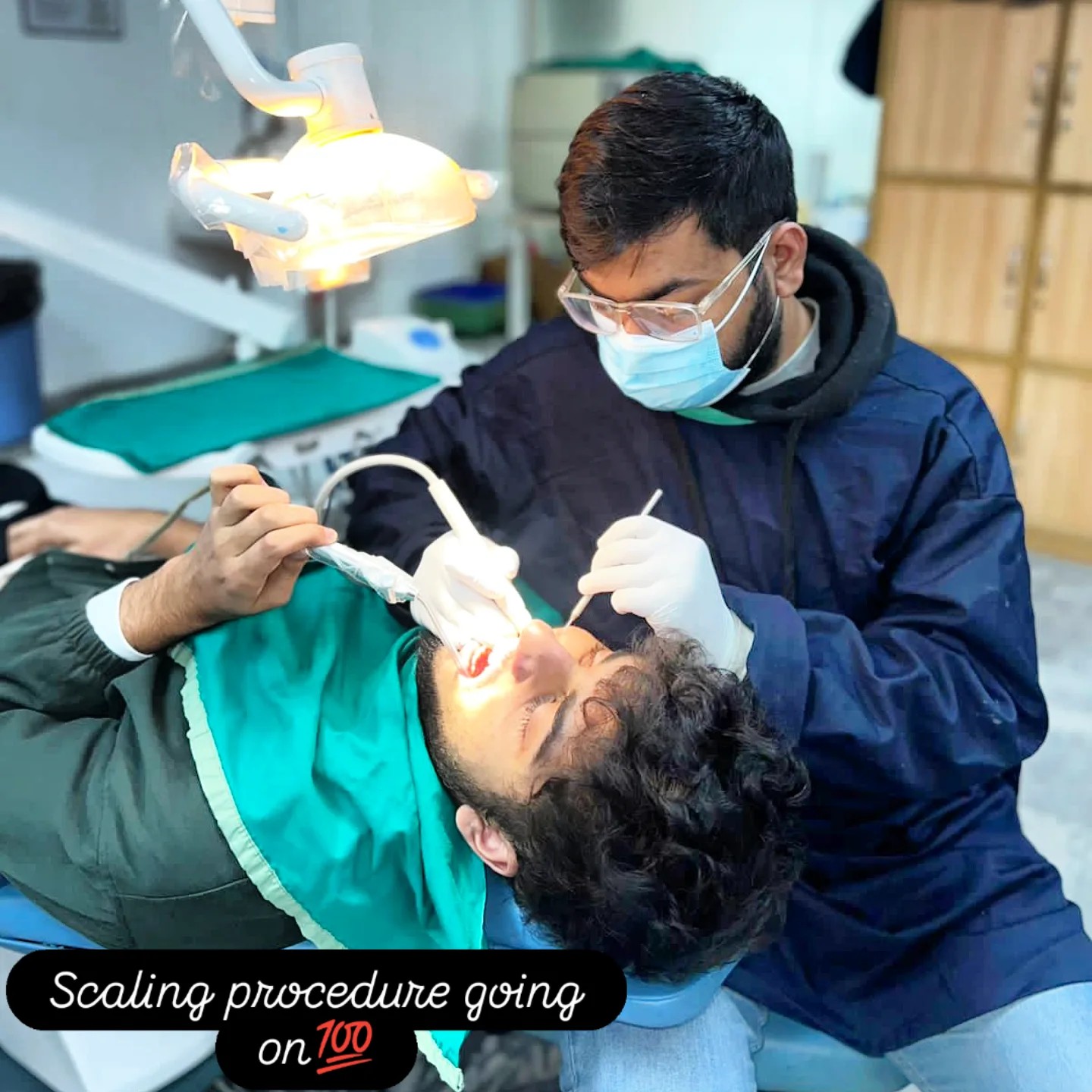
Routine six-monthly check. We took a bite-wing X-ray of the filling and the adjacent contact to confirm no recurrent caries. The margin was clean. A routine scaling was performed at the same visit.
Dr. Mian Momin Ahmad
“A Class II filling at the reversible-pulpitis stage is one of the most satisfying cases in general dentistry. The patient comes in with a pattern that has a clear diagnosis, a clear treatment, and a clear timeline. The work is technically careful but not technically dramatic. And the patient walks out — usually the same afternoon — eating normally. Cases caught at this stage are also cases that would have required a root canal six months later. Timing matters.”
Six habits that protect a Class II filling for a decade.
A filling lasts as long as the cavity does not recur at its margins. These six habits are what we asked Qari Sultan to commit to.
Floss daily — particularly at the new contact point
The single most important habit for protecting a Class II filling is daily flossing at the proximal contact. A waxed dental floss, slid through the contact and drawn carefully against the side of the new restoration once a day, removes the plaque that is the leading cause of recurrent caries at the margins. Ten seconds per tooth, every night.
Brush thoroughly twice a day with a fluoride toothpaste
A regular fluoride toothpaste — Sensodyne, Colgate Total — used with a soft-bristled brush for two minutes morning and night is the right baseline. The fluoride helps remineralise any micro-demineralisation that might otherwise lead to a new lesion forming on the same tooth.
Reduce snacking frequency, not just sugar amount
It is not the total quantity of sugar that causes cavities. It is the frequency with which the teeth are exposed to it. Six small sweet teas across a working day cause more acid exposure than one large dessert at dinner, even if the total sugar is similar. We asked Qari Sultan to consolidate his sweetened tea breaks into two or three sittings rather than spreading them across the day.
Come back if you feel any new tenderness in the same tooth
In the first 48 hours after a filling, some mild tenderness on biting is normal as the bite settles. After 48 hours, the tooth should feel like nothing at all. Any new tenderness developing later is worth checking. The fix is usually a five-minute bite adjustment with articulating paper, not a re-do of the filling.
Avoid very sticky foods on the new filling for the first 24 hours
Composite resin reaches full polymerised strength within minutes of curing, so the filling is functionally durable from the time you walk out of the clinic. But for the first 24 hours, the bonding interface is still settling at the molecular level, and very sticky or chewy foods can occasionally compromise the early seal. After the first day, no food restrictions apply.
Six-monthly recall, always
Every filling patient at our clinic comes back at six months for a marginal check. The check includes a visual inspection of the proximal surface, a probing of the gum at the cervical margin of the restoration, and a bite-wing X-ray every 18 months to confirm there is no recurrent caries forming under the margin. Twenty minutes per recall.
The window between "a filling" and "a root canal" is sometimes only months.
Pain that comes on with food and ends shortly afterward is one of the most informative symptoms in dentistry. It almost always means a cavity has reached the dentine but not yet damaged the pulp irreversibly. At this stage the treatment is a filling — about 70 minutes of chair time, a single visit, and a return to normal eating the same day.
Qari Sultan came in at the right time. The case was a single-visit filling. Waiting another six months would likely have meant a multi-visit root canal instead. The earlier the intervention, the smaller the procedure.
More on fillings, decay, and pulpitis.
More composite filling cases.
Every case in this archive is a real Odonto patient with their written consent.
Pain on chewing that comes and goes? Let's look at it.
The first 15 minutes are free. We will examine the affected tooth, take any X-rays needed, and tell you honestly whether the case is at the filling stage or the root-canal stage. There is no pressure to start treatment the same day.