One tooth, two cavities, one visit — Ms. Warda's bilateral fix.
Ms. Warda is a 27-year-old patient who came in to our Engineers Town clinic with two complaints that turned out to be one diagnosis — hot and cold sensitivity in a single tooth, and food impaction on both sides of that same tooth. This is the case file for the single-visit MOD composite restoration that resolved both surfaces of bilateral Class II caries in 90 minutes.
 Before
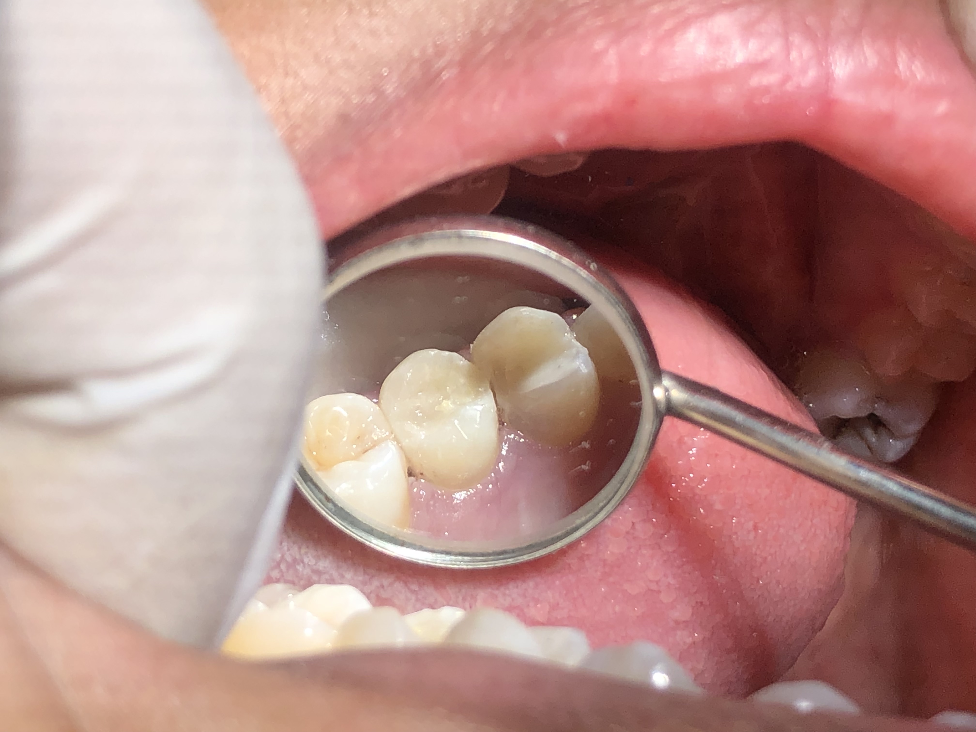 After
AfterTwo complaints. One unusual but textbook diagnosis.
Most adult composite fillings address a single carious lesion on a single surface. Ms. Warda's case is a useful example of how a single tooth can develop parallel decay on both proximal surfaces when the original contact anatomy was slightly under-contoured.
Ms. Warda came in to our clinic on a weekday morning. She is approximately 27 years old, a working professional, and a patient who had been quietly managing two recurring annoyances for several months. She described them precisely. First, she felt a sharp sensitivity in one tooth on her right side whenever she drank tea or anything cold from the refrigerator. The sensitivity was localised — she could point to it — and it peaked quickly and resolved within thirty seconds of stopping. Second, she was getting food stuck between her teeth in the same area at every meal. Particularly meat and stringy vegetables. She had been carrying floss in her purse for several months as a routine after-meal habit.
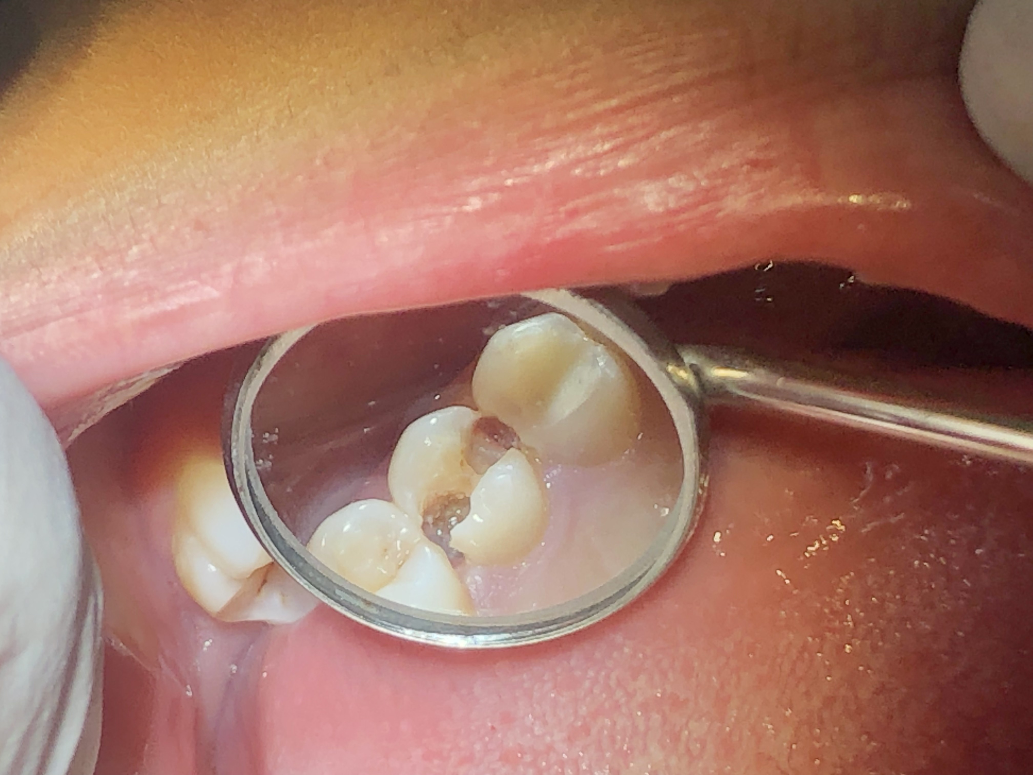
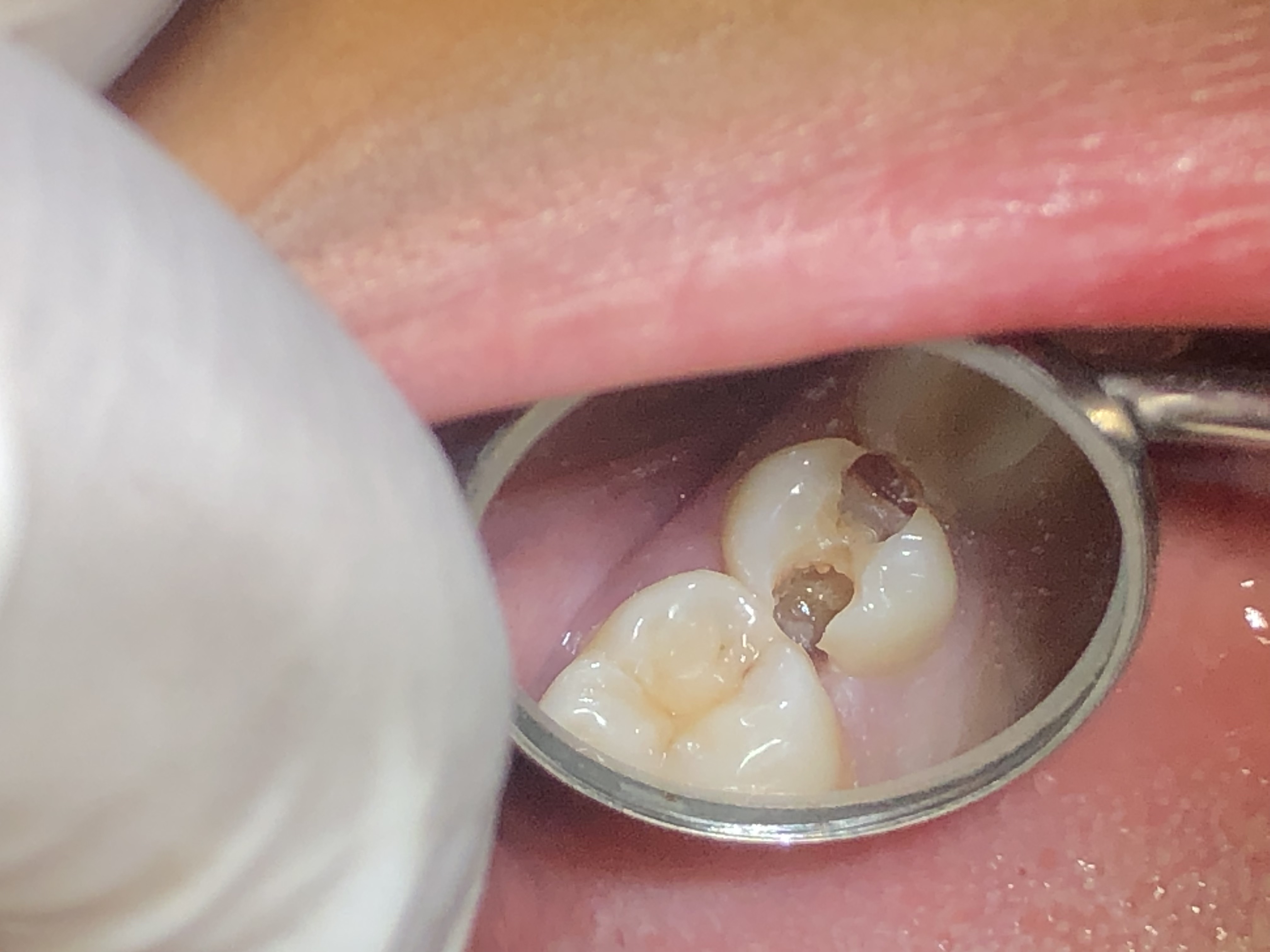
The clinical examination took 20 minutes. We took an intra-oral photograph of the affected quadrant, a periapical X-ray, and — most usefully — a bite-wing X-ray of the right side. The bite-wing image showed both lesions side-by-side on the same tooth. Bilateral Class II caries on the mesial and distal proximal surfaces of the same posterior tooth. Both lesions extended into the dentine. Neither had reached the pulp.
The cold-stimulus test on the affected tooth produced a sharp, brief response that ended within five seconds of removing the stimulus. The two adjacent teeth tested cleanly. The periapical bone was healthy. The diagnosis was clear: bilateral Class II caries with reversible pulpitis. The treatment was a single-visit MOD composite restoration.
We walked Ms. Warda through what we had found, with the bite-wing X-ray on the screen. We pointed out the two carious lesions, explained why a single tooth had developed parallel decay on both sides (a slightly under-contoured contact anatomy combined with frequent snacking), and described what the restoration would involve. We discussed the option of splitting the work into two visits — one side first, the other side later — and recommended doing both in a single sitting, both because it was safer (no temporary fillings to leak) and because the patient would only need to be numbed once.
She agreed, we numbed the quadrant, and the procedure took 90 minutes start to finish. She walked out the same morning with both proximal surfaces restored, the food impaction gone, and the sensitivity already noticeably reduced. By the second day post-treatment, all sensitivity had resolved.
There is a detail worth dwelling on in Ms. Warda's case because it is one of the most under-discussed elements of routine restorative dentistry — the under-contoured proximal anatomy that allowed the bilateral caries pattern to develop in the first place. When a posterior tooth has slightly flat proximal surfaces — rather than the gently bulged shape that a healthy contact point creates — food packs into the embrasures at every meal. Plaque accumulates more quickly than the toothbrush can clear it. The pH at the contact point drops repeatedly through the day, demineralising the enamel surface micrometre by micrometre. Over 18 to 24 months, the demineralisation crosses the enamel-dentine junction and the lesion becomes a true cavity. The new MOD restoration we built for Ms. Warda restored the proper proximal contour on both sides, with snap-fit contacts that now deflect food the way a healthy tooth always should have.
This contour restoration is the part of an MOD case that takes the most time and the most care, and it is the part most directly responsible for whether the case lasts ten years or twenty. We used a sectional matrix system that produces a slight outward curve of the proximal wall — imitating the natural bulge of a healthy contact. We built each proximal wall in 2 mm increments, curing each layer for 20 seconds, so that the polymerisation shrinkage of the composite was distributed across many small increments rather than concentrated in a single bulk fill. We checked the contact with floss at the end of placement — the floss had to snap through with the same resistance as the contacts between her healthy neighbouring teeth. Every step had a measurable check at the end.
The other technical detail that matters here is the laser-assisted excavation we used on the cavity floors. The soft-tissue diode laser produces a brief pulse of energy that disinfects the dentine surface as the rotary bur exposes it. The published evidence for laser-assisted disinfection in cavity floors is modest but consistent: a small reduction in post-operative sensitivity, a small improvement in the long-term seal of the restoration. For an MOD case, where there are two cavity floors rather than one, the benefit is approximately doubled. Both of Ms. Warda's cavity floors received a laser disinfection pass before bonding, and her near-zero post-operative sensitivity is consistent with the expected pattern.
By the time the dam was removed and the bite was checked, Ms. Warda had a tooth with two new proximal walls that snap-fit-flossed correctly, an occlusal anatomy that matched the rest of her dentition, a polish that brought the composite to a high gloss matching her natural enamel, and a written aftercare sheet in her hand. She walked out the same morning. She messaged at 8 PM that evening to confirm she had eaten dinner without any food getting stuck — for the first time in many months.
Four findings — two cavities, one diagnosis.
The bilateral pattern is unusual but well-understood. Once we had confirmed there was no deeper pulpal involvement, the treatment plan was straightforward.
Bilateral Class II caries — both proximal surfaces of the same tooth
When we examined Ms. Warda's posterior tooth carefully with a fine dental explorer and confirmed our findings with a bite-wing X-ray, the pattern was unusual but not rare. The affected molar had carious lesions on both of its proximal surfaces — the mesial side (facing the neighbouring tooth in front) and the distal side (facing the tooth behind). Both lesions were of comparable depth, both extended into the dentine, and both were responsible for trapping food at the contact points on either side of the tooth. The pattern usually develops over time when a single tooth has slightly under-contoured proximal contacts on both sides — food packs in routinely, plaque accumulates, and decay starts at both surfaces in parallel.
Hot and cold sensitivity localised to the affected tooth
Ms. Warda described a sharp sensitivity when she drank tea or anything cold from the fridge, and the sensitivity was clearly localised to one tooth on her right side. The sensitivity peaked within a few seconds of the temperature change and resolved within about thirty seconds of the stimulus being removed. This pattern of brief, sharp, stimulus-provoked sensitivity is consistent with reversible pulpitis — the pulp is irritated by the proximity of the caries but has not been damaged irreversibly.
Persistent food impaction in the affected area
The second part of her complaint was a constant sense of food getting stuck between her teeth in the same area — particularly meat and stringy vegetables. Every meal required her to use a toothpick or floss afterwards to dislodge what had packed into the proximal embrasures on both sides of the tooth. This food impaction was both a quality-of-life complaint and a clinical sign — the under-contoured contacts created by the carious lesions were no longer functioning as proper food deflectors.
No deeper pulpal involvement on cold testing
A cold-stimulus test on the affected tooth produced a sharp, brief response that ended within five seconds of removing the stimulus. The two neighbouring teeth tested cleanly. The periapical radiograph showed normal periapical bone around the root tips with no widening of the periodontal ligament space. The pulp was healthy. A two-surface restoration would resolve the entire problem.
Four phases. All in one 90-minute sitting.
Doing both surfaces in one visit is safer and more comfortable than splitting them into two visits with temporary fillings in between. The whole procedure was completed under a single rubber-dam isolation.
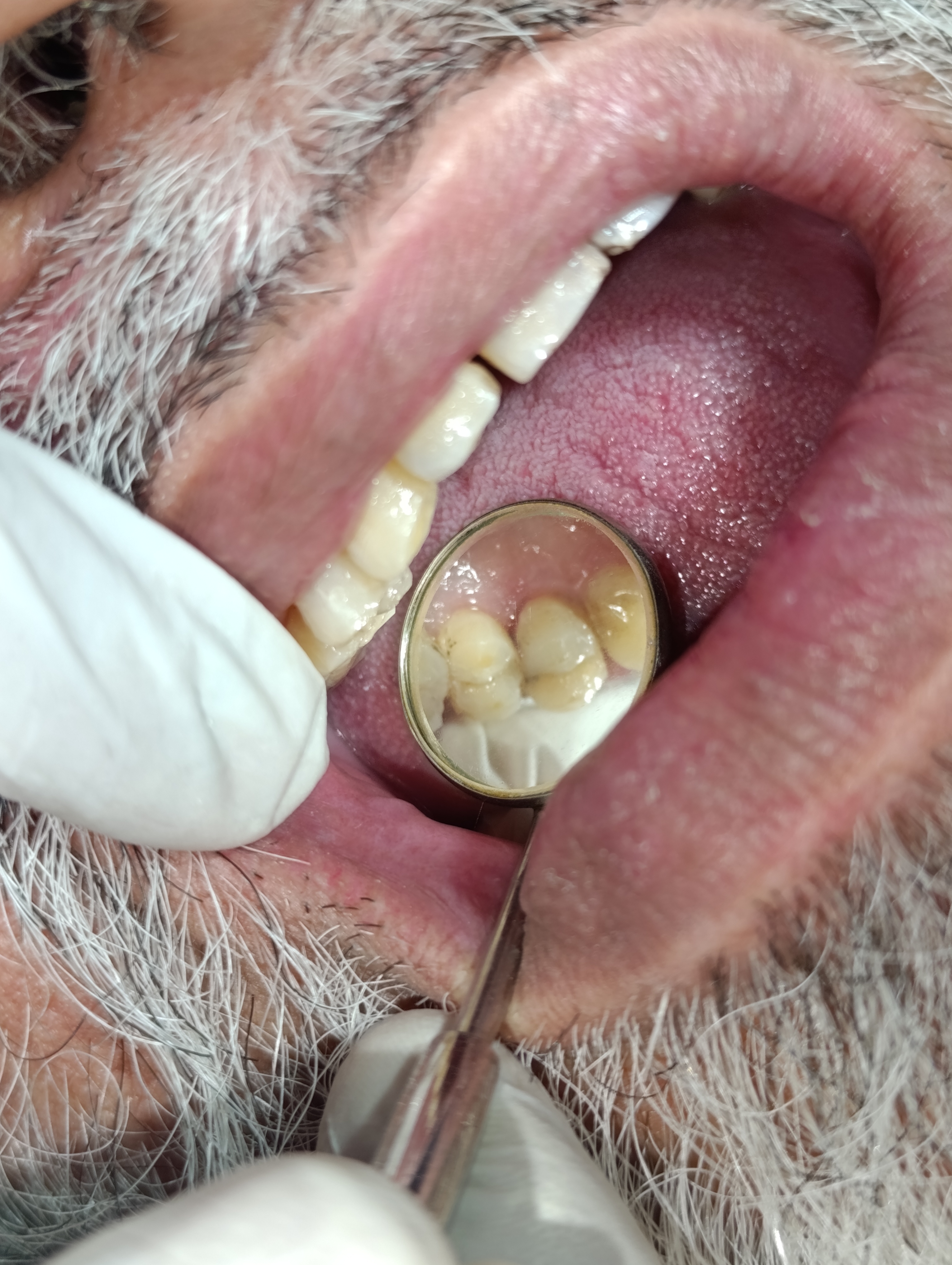
Examination, photographs and bite-wing X-ray
We took a focused intra-oral photograph of the affected quadrant, a bite-wing X-ray, and a periapical X-ray for confirmation. The bite-wing was the key image — it showed both proximal lesions on the same tooth simultaneously, allowing us to plan the restoration as a single mesial-occlusal-distal (MOD) procedure rather than two separate fillings. We performed a cold-test on the affected tooth and the two adjacent teeth to confirm pulpal status.
Phase 1 · ~ 15 minLocal anaesthesia and rubber dam isolation
A buccal infiltration of 4% articaine, supplemented by a single palatal injection, gave complete anaesthesia of the working tooth and surrounding tissues. We placed a rubber dam over the affected tooth and both adjacent teeth, with a sectional matrix system pre-placed to receive the composite. Rubber-dam isolation for an MOD restoration is especially important — saliva contamination at either proximal margin would compromise the bond on that side.
Phase 2 · ~ 15 minBilateral laser-assisted caries excavation
We removed the carious dentine from the mesial cavity first using a sequence of round burs and finished it with a laser disinfection pass. We then turned attention to the distal cavity and repeated the same sequence. Both cavity floors were checked with a caries-detector dye to confirm complete removal of infected dentine. The healthy remaining dentine on both sides was etched with phosphoric acid, rinsed, and dried, ready for bonding.
Phase 3 · ~ 30 minSequential composite layering, sculpting and polish
We built up the mesial wall first using a sectional matrix, layering the composite in 2 mm increments and curing each layer for 20 seconds. We then re-positioned the matrix to the distal side and built the distal wall the same way. Once both proximal walls were complete, we filled the central occlusal space and sculpted the final anatomy. A high-speed diamond bur refined the marginal ridges, and a sequence of polishing wheels brought the final surface to a high gloss. Floss was used to confirm snap-fit contacts on both sides before the dam was removed.
Phase 4 · ~ 40 minSame patient. Same tooth. Same day.
Drag the divider across the photo to compare. Both photos were taken in our Lahore clinic — "before" at the diagnostic visit, "after" immediately following the polish.
BeforeAfterWhy an MOD is technically harder than a single-surface filling.
A mesial-occlusal-distal restoration is one of the most technique-sensitive procedures in routine restorative dentistry. Three things have to be done correctly for the restoration to last.
Two separate proximal contacts have to be re-created
Each proximal contact must be tight enough to prevent food impaction but not so tight that floss cannot pass through. We use a sectional matrix that is placed on one side at a time and provides a slight outward pressure on the proximal wall — recreating the natural bulge of a healthy contact. Doing this twice on the same tooth requires placing, building, removing, and re-placing the matrix on the opposite side, all within the same anaesthetic and the same rubber-dam isolation.
The remaining tooth structure has to stay strong enough to chew on
An MOD restoration removes more tooth structure than a single-surface filling and weakens the cusps proportionally. We compensate by using a slightly tougher composite material and by building the restoration in incremental layers that distribute the polymerisation shrinkage across many smaller, lower-stress increments. The final cusp shape is sculpted to be very slightly out of strong contact in protrusive movements — this reduces the lateral load on the cusps over years of chewing.
Both cavity floors are checked under magnification for complete excavation
A residual pocket of infected dentine left at the floor of either cavity will lead to recurrent caries under the filling within a few years. We use a caries-detector dye that stains residual infected dentine, allowing us to confirm complete excavation before bonding begins. The laser disinfection pass reduces the residual bacterial load further. Together these two steps make recurrent caries within the first decade extremely unlikely.
Five questions we hear at every MOD consult.
These are the worries we heard from Ms. Warda and the worries we hear from most patients facing a two-surface posterior filling. Tap any one to read the long answer.
Why did one tooth develop cavities on both sides at the same time?+
This is a question worth answering carefully because it has implications for the home-care plan going forward. A single tooth developing carious lesions on both proximal surfaces simultaneously almost always points to a contact-quality problem — the contact points on either side of the tooth were slightly under-contoured to begin with, allowing food to pack into both embrasures routinely.
For Ms. Warda, the original anatomy of the affected tooth had slightly flat proximal surfaces rather than the gently bulged shape that deflects food away from the embrasure. Combined with her snacking pattern — multiple small sweet teas across the working day — the persistent plaque accumulation on both sides led to parallel demineralisation on both proximal surfaces. The result, after about 18 to 24 months of slow progression, was the bilateral pattern we found at diagnosis.
The new restoration restores the proper proximal contour on both sides, with snap-fit contacts that deflect food the way the original tooth should have. Combined with a slightly more disciplined flossing routine and reduced snacking frequency, this should prevent the same pattern from recurring on neighbouring teeth.
Will my hot/cold sensitivity disappear completely after the filling?+
Yes — almost always, and for predictable reasons. The sensitivity you were experiencing was caused by the close proximity of the cavity to the pulp. As the carious lesion progresses through dentine, the dentinal tubules near the pulp transmit hot and cold stimuli more rapidly than they would through healthy enamel. Removing the carious tissue and sealing the cavity with a bonded composite restoration restores the normal insulation of the dentine.
In the first 24 to 48 hours after a deep filling, some mild residual sensitivity to cold is normal as the bonded interface settles. After that window, the sensitivity should be completely gone. For Ms. Warda, the sensitivity resolved by the second day post-treatment and has not recurred at her six-month follow-up.
If sensitivity ever returns to the same tooth weeks or months later, that is something to flag immediately. It can occasionally indicate either a marginal failure of the filling or a deeper pulpal issue that is developing. Both are addressable if caught early.
Is it safe to do both fillings in one visit?+
For an MOD (mesial-occlusal-distal) restoration on a single tooth, doing both surfaces in one visit is not just safe — it is the standard of care. Splitting the procedure into two visits would mean placing a temporary filling on one side for several weeks, with potential for marginal leakage and temporary food impaction in the intervening period.
The technical considerations for a single-visit MOD are about the sequencing of the work, not about safety. We use a sectional matrix system that can be placed first on one proximal side, then re-positioned to the other, allowing each proximal wall to be built up separately under proper contour control. The whole procedure takes about 70 to 90 minutes of chair time, with continuous local anaesthesia. The patient is comfortable throughout.
For Ms. Warda, the entire bilateral procedure was completed in a single 90-minute appointment with no breaks, no complications, and no need for a second visit.
How is this different from a crown? Should I have had a crown instead?+
A crown is indicated when a tooth has lost so much structure that a filling can no longer be reliably retained or when the remaining cusps are likely to fracture under chewing load. Both situations are common after very large cavities or after root canal treatment.
For Ms. Warda, neither situation applied. The two carious lesions, although on opposite sides, were both relatively small to moderate in size. The cusps were not undermined. The remaining tooth structure was strong enough to retain a bonded composite restoration without any cuspal coverage. A crown would have meant removing significantly more tooth structure than the restoration actually required.
We always favour the smallest restoration that adequately solves the problem. For Ms. Warda's tooth, that was a bonded MOD composite. If the same tooth had presented with cuspal undermining or pulpal involvement requiring root canal treatment, the answer would have been a crown — but not on these clinical findings.
How can I prevent the same pattern of decay on my other teeth?+
This is the question that determines whether Ms. Warda's next ten years will involve more fillings or none at all, and it deserves a detailed answer. The bilateral Class II pattern she had developed was driven by two factors: a slightly under-contoured original proximal anatomy on the affected tooth, and a snacking pattern that exposed her enamel to acidic conditions multiple times each day. The new restoration has addressed the first factor. The second is up to her to manage.
The cariogenic effect of sugar is determined more by frequency of exposure than by total quantity. A patient who drinks six small sweet teas across the working day exposes their teeth to about six episodes of acidic pH lasting roughly 30 minutes each — three hours of acid exposure spread across the day. The same patient drinking one large sweet drink at dinner exposes their teeth to a single 30-to-45-minute episode. Same total sugar; very different cariogenic profile. We asked Ms. Warda to consolidate her sweetened tea breaks into two or three sittings rather than six or seven.
The second prevention measure is mechanical: nightly flossing at the proximal contacts of every posterior tooth, not just the recently treated one. A waxed dental floss, slid through each contact and drawn carefully against both adjacent teeth, takes about 90 seconds to complete the entire posterior dentition. Done every night before brushing, it removes the plaque that the toothbrush cannot reach and prevents the development of a new lesion at any proximal site.
The third measure is the fluoride exposure of her toothpaste. A regular fluoride toothpaste at the standard 1450 parts-per-million concentration, used for two minutes morning and night, helps remineralise any micro-demineralisation that might otherwise lead to a new lesion. We asked her not to rinse aggressively after brushing — letting a small residual film of fluoride remain on the teeth for a few minutes extends the remineralisation benefit.
With these three habits in place, the realistic expectation is that no new caries lesions will develop in her posterior dentition for at least the next ten years, and possibly indefinitely.
How much did this cost?+
A bilateral Class II MOD restoration is charged at our two-surface composite filling rate, which includes the consultation visit if it leads directly to treatment, the radiographs, the local anaesthesia, the rubber-dam isolation, the laser-assisted excavation on both sides, the layered composite placement, and the final polish. There is no additional charge for the laser use, the rubber dam, or the bite-wing X-ray.
The fee is quoted in writing at consultation. For Ms. Warda, the entire procedure was completed within a single all-inclusive fee. No add-ons appeared on the final invoice that were not on the original written quote.
For a comparable case, we generally quote our two-surface MOD composite at the same rate regardless of whether the surfaces are on the same tooth or on adjacent teeth, because the working time and technical effort are comparable.
The follow-up visits.
A single-visit MOD restoration is followed by a one-week review and routine six-monthly recall. Here is how Ms. Warda's follow-up went.
She messaged on the second day post-treatment to confirm the cold sensitivity had completely resolved. No new pain. Both proximal contacts felt snap-fit during flossing.
A short 10-minute visit to check the bite with articulating paper. The occlusion was already settled — no high spots to adjust. The proximal contacts on both sides remained snap-fit. She reported zero food impaction since the procedure.
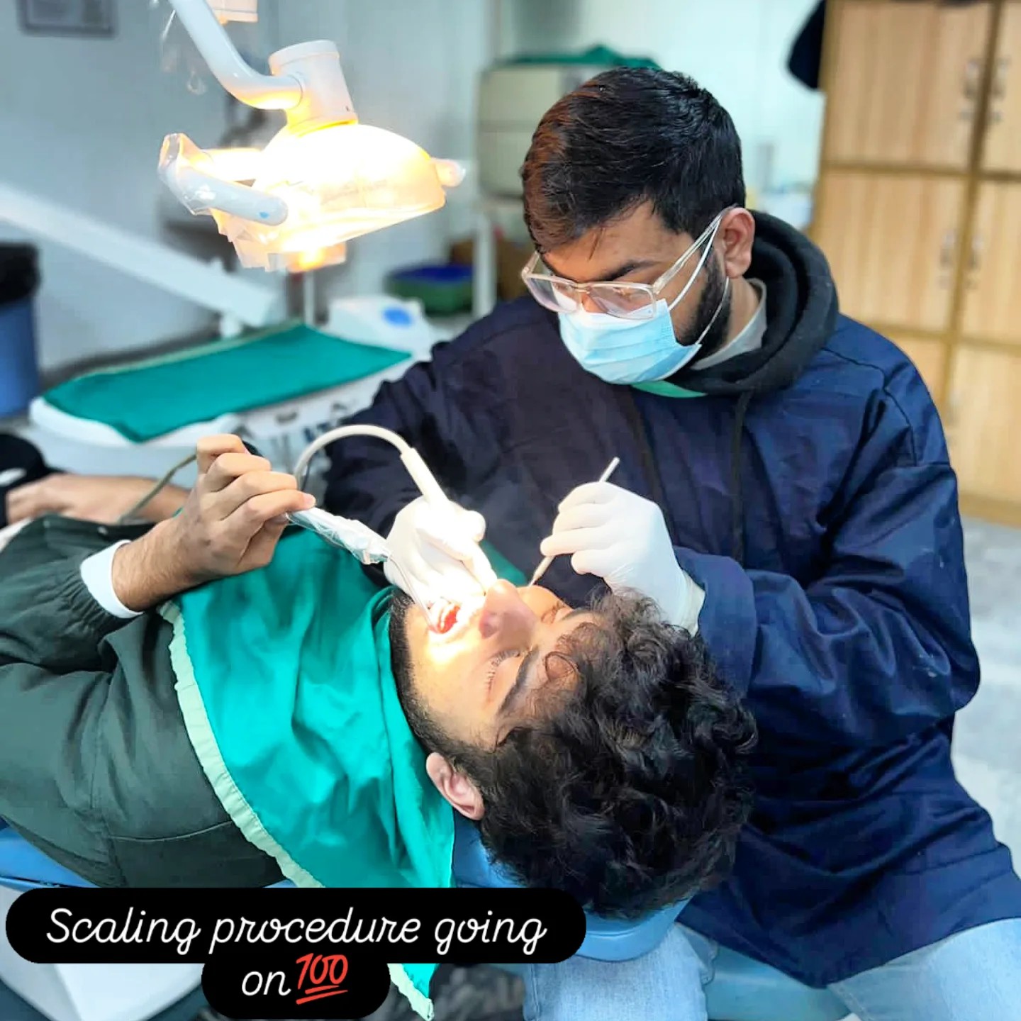
Routine six-monthly check. Bite-wing X-ray confirmed no recurrent caries forming under either proximal margin. The proximal contacts were intact. A routine scaling was performed at the same visit.
Dr. Mian Momin Ahmad
“Bilateral Class II caries on the same tooth is uncommon but not rare, and when it does occur it usually points to a slightly under-contoured proximal anatomy from the start. Treating it well in a single visit requires careful matrix work and incremental layering, but the procedure is fundamentally the same as a single-surface filling — just done twice in the same anaesthetic. Patients like Ms. Warda are a reminder that small problems can pile up quietly until they cross a threshold, and that the threshold is usually crossed at the dinner table.”
Six habits that protect an MOD restoration.
A two-surface filling has two sets of margins, two proximal contacts, and twice the surface area where recurrent caries could start. The aftercare regimen reflects that.
Floss carefully through both new proximal contacts
Both contacts on either side of the new filling need to be flossed every night. Waxed dental floss, slid through the contact and drawn carefully against the side of the new restoration once a day, removes the plaque that is the leading cause of recurrent caries. Ten seconds per contact, both sides, every night. For Ms. Warda this is the single most important habit going forward.
Watch for renewed food impaction
Properly contoured proximal contacts deflect food away from the embrasure. If she ever notices food packing into the same areas the way it did before treatment, that may be an early sign of contact loosening — usually fixable with a small contour adjustment, occasionally requiring a small repair of the proximal margin. We asked her to message us on WhatsApp if she ever noticed this.
Reduce snacking frequency
Decay risk is driven by the frequency of sugar exposure more than the total quantity. Spreading sweet drinks across the working day causes more enamel acid exposure than the same total at one sitting. We asked Ms. Warda to consolidate her sweetened tea breaks into two or three sittings rather than the six or seven she had been having.
Brush twice a day with a fluoride toothpaste
A regular fluoride toothpaste used with a soft-bristled brush for two minutes morning and night is the baseline. The fluoride helps remineralise any micro-demineralisation that might otherwise lead to a new lesion forming on a neighbouring tooth.
Be cautious with the new filling for the first 24 hours
Composite resin reaches full strength within minutes of curing, so the filling is functionally durable from the time of walking out of the clinic. But for the first 24 hours we asked her to avoid very hard or very sticky foods on the working side — chewing gum, hard caramel, raw carrots — to allow the bonded interface to settle without stress.
Six-monthly recall with bite-wing X-ray every 18 months
Every filling patient at our clinic comes back at six months for a marginal check. The check includes a visual inspection, gum probing at the cervical margin, and a bite-wing X-ray every 18 months to confirm there is no recurrent caries forming under the proximal margins. Twenty minutes per recall.
Food impaction at the same site is rarely a coincidence.
Patients who notice food packing between the same teeth at every meal often assume it is a quirk of their anatomy that they have to live with. Sometimes it is. Often it is the earliest functional sign of a developing cavity at the contact point, before any pain has begun. Catching the case at this stage usually means a single filling resolves it; missing it for another year often means two fillings or a more complex procedure.
Ms. Warda came in at the right stage. The case was a single-visit two-surface filling. Earlier intervention is almost always smaller intervention in restorative dentistry.
More on fillings, caries, and contact restoration.
More composite filling cases.
Food getting stuck in the same place every meal? Let's check it.
The first 15 minutes are free. We will examine the affected area, take a bite-wing X-ray if needed, and tell you honestly whether the case is at the filling stage or something more. There is no pressure to start treatment the same day.